Alternate header for print version
Contributors
Help
Submit
Search
menu
Data sets
Videos
Latest data
Center for Research in Biological Systems
Basic Science Building, Room 1000
University of California, San Diego
9500 Gilman Drive
La Jolla, CA 92093-0608, USA
Voice
: (858) 534-0276
Fax
: (858) 534-7497
Email
: dorloff@ncmir.ucsd.edu
Search Results for
Rattus
(571 results)
CIL:11385
NCBI Organism Classification
Rattus
Biological Process
post-translational protein modification
Cellular Component
Golgi apparatus
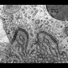
Figure 215 (upper) and 216 (higher magnification, lower) from Chapter 6 (Golgi Apparatus) of 'The Cell, 2nd Ed.' by Don W. Fawcett M.D. Granules of the eosinophilic myelocyte from rat bone marrow sta...
CIL:11486
NCBI Organism Classification
Rattus
Biological Process
cellular catabolic process
Cellular Component
peroxisome
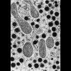
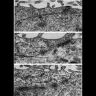
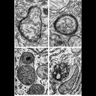
Figures 282 (upper) and 283 (lower) from Chapter 9 (Peroxisomes) of 'The Cell, 2nd Ed.' by Don W. Fawcett M.D. Examples longitudinal, or transverse sections through peroxisomes from rat liver hepatoc...
CIL:11199
NCBI Organism Classification
Rattus
Biological Process
cell-cell adhesion
Cellular Component
zonula adherens
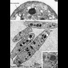
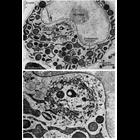
These examples of ependymal epithelium from the rat brain represent epithelia that lack zonulae occludentes. Further, there appears to be no consistent order to the junctional specializations that are...
CIL:10841
NCBI Organism Classification
Rattus
Biological Process
autophagy
Cellular Component
autophagic vacuole
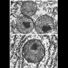
Figures 266 (upper left), 267 (upper right), 268 (lower left) and 269 (lower right) from Chapter 8 (Lysosomes) of 'The Cell, 2nd Ed.' by Don W. Fawcett M.D. Autophagic vacuoles from normal rat liver ...
CIL:11933
NCBI Organism Classification
Rattus
Biological Process
G1/S transition of mitotic cell cycle
Cellular Component
mitochondrion
Rat NRK stably expressing RFP targeted to mitochondrial matrix imaged at the cell cycle stage G1-S transition. This image is a z-stack from a living cell collected on a Zeiss 510 LSM confocal using a ...
CIL:23041
NCBI Organism Classification
Rattus
Biological Process
gelsolin treatment
Cellular Component
intermediate filament
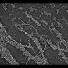
Association of plectin with myosin II. A gelsolin-treated REF-52 cytoskeleton immunogold labeled myosin II (10 nm). After actin depletion, intermediate filaments with plectin sidearms remain associate...
CIL:36052
NCBI Organism Classification
Homo sapiens
Biological Process
microtubule cytoskeleton organization
Cellular Component
microtubule
Figures 417 (upper) and 418 (lower) from Chapter 16 (Cytoplasmic matrix and cytoskeleton) of 'The Cell, 2nd Ed.' by Don W. Fawcett M.D. A marginal bundle of microtubules is associated with the ellipti...
CIL:35979
NCBI Organism Classification
Rattus
Biological Process
lipid storage
Cellular Component
lipid particle
Figure 357 from Chapter 15 (Cytoplasmic Inclusions) of 'The Cell, 2nd Ed.' by Don W. Fawcett M.D. Intestinal epithelium of rat in an advanced stage of fat absorption. In contrast to most cells, lipi...
CIL:35996
NCBI Organism Classification
Rattus
Biological Process
secretory granule organization
Cellular Component
secretory granule
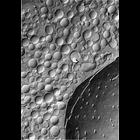
Figure 375 from Chapter 15 (Cytoplasmic Inclusions) of 'The Cell, 2nd Ed.' by Don W. Fawcett M.D. Freeze-fracture replica of a cell from rat adrenal medulla. Using this technique, secretory vesicles...
CIL:37168
NCBI Organism Classification
Rattus
Biological Process
adherens junction organization
Cellular Component
adherens junction
Transmission electron micrograph of adherens junctions (macula adherens or desmosome)in the proximal tubule of a rat kidney. The junctions with associated tonofilaments are seen at the apposed plasma...
« Previous
1
...
6
7
8
9
10
11
12
13
...
58
Next »
Results per page:
10
20
50
100

")