Alternate header for print version
Contributors
Help
Submit
Search
menu
Data sets
Videos
Latest data
Center for Research in Biological Systems
Basic Science Building, Room 1000
University of California, San Diego
9500 Gilman Drive
La Jolla, CA 92093-0608, USA
Voice
: (858) 534-0276
Fax
: (858) 534-7497
Email
: dorloff@ncmir.ucsd.edu
Search Results for
Rattus
(571 results)
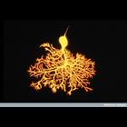
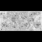
CIL:39010
NCBI Organism Classification
Rattus
Biological Process
cerebral Purkinje cell morphogenesis
Cellular Component
neuronal cell body
Confocal microscope image of a developing Purkinje cell in the cerebellum of a 9 day postnatal rat. The cerebellum contains millions of these cells which are involved with integrating motor and sensor...
CIL:40666
NCBI Organism Classification
Rattus
Biological Process
dendritic spine organization
Cellular Component
dendritic spine
Platinum replica illustrating the cytoskeletal organization of dendritic spines from extracted 14 DIV neurons. This image shows branched actin filaments (cyan) in the neck of respective spines. This i...
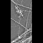
CIL:40667
NCBI Organism Classification
Rattus
Biological Process
dendritic spine organization
Cellular Component
dendritic spine
Platinum replica illustrating the cytoskeletal organization of dendritic spines from extracted 14 DIV neurons. Thin spines associate with dendrites at the base (bottom) and with axons by the head (top...
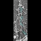
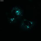
CIL:40967
NCBI Organism Classification
Rattus
Biological Process
hippocampus development
Cellular Component
neurofilament cytoskeleton
Widefield multiphoton fluorescence image of Rat hippocampus stained to reveal the distribution of glia (cyan), neurofilaments (green) and cell nuclei (yellow). 2nd Prize, 2010 Olympus BioScapes Digit...
CIL:9153
NCBI Organism Classification
Rattus
Biological Process
none specified
Cellular Component
synapse
EM micrograph demonstrating the colocalization of GABA neurotransmitter (30 nm gold) and delta 1/2 glutamate receptors (10 nm gold) in the cerebellar cortex molecular layer at P10. It shows Delta labe...
CIL:9971
NCBI Organism Classification
Rattus
Biological Process
constitutive secretory pathway
Cellular Component
Golgi apparatus
Normal Rat Kidney (NRK) cells grown in culture expressing Galactosyl Transferase-YFP (GalT-YFP) and p58-CFP. This file is the CFP time series demonstrating the organization of the early secretory path...
CIL:11364
NCBI Organism Classification
Rattus
Biological Process
post-translational protein modification
Cellular Component
Golgi apparatus
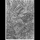
Figure200 from Chapter 6 (Golgi Apparatus) of 'The Cell, 2nd Ed.' by Don W. Fawcett M.D. The Golgi apparatus in epithelial cells of rat epididymis. The tissue was stained with thiamine pyrophosphata...
CIL:11374
NCBI Organism Classification
Rattus
Biological Process
post-translational protein modification
Cellular Component
Golgi apparatus
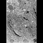
Figure 208 from Chapter 6 (Golgi Apparatus) of 'The Cell, 2nd Ed.' by Don W. Fawcett M.D. Golgi apparatus and associated organelles in a rat liver hepatocyte. The star indicates a region of continui...
CIL:11376
NCBI Organism Classification
Rattus
Biological Process
post-translational protein modification
Cellular Component
Golgi apparatus
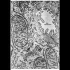
Figure 209 from Chapter 6 (Golgi Apparatus) of 'The Cell, 2nd Ed.' by Don W. Fawcett M.D. Portions of two hepatic cells and the intervening bile canaliculus from rat liver. Two stacks of Golgi ciste...
CIL:11384
NCBI Organism Classification
Cavia porcellus
Biological Process
post-translational protein modification
Cellular Component
Golgi apparatus
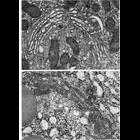
Figures 213 (upper) and 214 (lower) from Chapter 6 (Golgi Apparatus) of 'The Cell, 2nd Ed.' by Don W. Fawcett M.D. The Golgi complex of a Leydig cell from guinea pig testis (upper) and from and acina...
« Previous
1
...
5
6
7
8
9
10
11
12
...
58
Next »
Results per page:
10
20
50
100


")