Alternate header for print version
Contributors
Help
Submit
Search
menu
Data sets
Videos
Latest data
Center for Research in Biological Systems
Basic Science Building, Room 1000
University of California, San Diego
9500 Gilman Drive
La Jolla, CA 92093-0608, USA
Voice
: (858) 534-0276
Fax
: (858) 534-7497
Email
: dorloff@ncmir.ucsd.edu
Search Results for
Rattus
(571 results)
CIL:37214
NCBI Organism Classification
Rattus
Biological Process
none specified
Cellular Component
nerve terminals
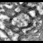
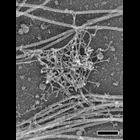
Transmission electron micrograph of nerve terminals from a rat tongue. Note that this very early (1953) micrograph is primarily of historical interest since it pre-dates glutaraldehye fixation which...
CIL:37215
NCBI Organism Classification
Rattus
Biological Process
none specified
Cellular Component
nerve terminals
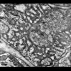
Transmission electron micrograph of nerve terminals from a rat tongue. Note that this very early (1953) micrograph is primarily of historical interest since it pre-dates glutaraldehye fixation which...
CIL:37219
NCBI Organism Classification
Rattus
Biological Process
transmission of nerve impulse
Cellular Component
myelin sheath
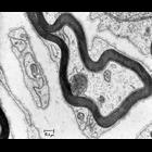
Transmission electron micrograph of an axon from a rat tongue surrounded by a myelin sheath. Image made available by James D. Jamieson and the Department of Cell Biology, Yale University School of M...
CIL:38985
NCBI Organism Classification
Rattus
Biological Process
cell adhesion
Cellular Component
stress fiber
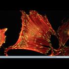
Fluorescent micrograph of rat embryo fibroblast cell growing in serum stained to reveal actin stress fibres (red) and vinculin (component of focal adhesions) in green/yellow.
CIL:41465
NCBI Organism Classification
Rattus
Biological Process
none specified
Cellular Component
neuron projection
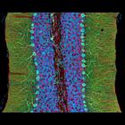
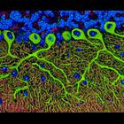
Photomicrograph of a midsaggital section showing the different components of the rat cerebellum, including Purkinje neurons in green, glia (non-neuronal cells) in red, and cell nuclei in blue. This i...
CIL:40804
NCBI Organism Classification
Rattus
Biological Process
dendrite morphogenesis
Cellular Component
axon
Colorized transmission electron micrograph of a platinum replica showing the cytoskeletal organization of stubby dendritic spines in extracted hippocampal neurons after 14 DIV. This image shows axons,...
CIL:40662
NCBI Organism Classification
Rattus
Biological Process
dendritic spine organization
Cellular Component
dendritic spine
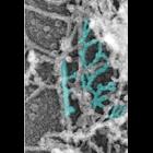
Platinum replica depicting the cytoskeletal organization of dendritic spines in extracted 14 DIV neurons. Branched actin filaments (cyan) in the head. Image corresponds to Figure 2a in Mol Biol Cell....
CIL:40663
NCBI Organism Classification
Rattus
Biological Process
dendritic spine organization
Cellular Component
dendritic spine
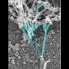
Platinum replica depicting the cytoskeletal organization of dendritic spines in extracted 14 DIV neurons. This image shows branched actin filaments (cyan) at the neck–head junction. Dashed arrows ...
CIL:41815
NCBI Organism Classification
Rattus
Biological Process
cerebellum structural organization
Cellular Component
inositol trisphosphate recepto
Midsaggital section of rat cerebellum, captured using confocal imaging. Section shows inositol trisphosphate receptor (IP3R) labelled in green, DNA in blue, and synaptophysin in magenta. Honorable Me...
CIL:47951
NCBI Organism Classification
Rattus
Biological Process
none specified
Cellular Component
primary cilium
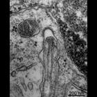
The electron micrograph shows the tip of a primary cilium in a testis cell in primary culture. The plasma membrane of the adjacent cell has a coated pit juxtaposed to the ciliary tip.
« Previous
1
2
3
4
5
6
7
8
9
...
58
Next »
Results per page:
10
20
50
100