Alternate header for print version
Contributors
Help
Submit
Search
menu
Data sets
Videos
Latest data
Center for Research in Biological Systems
Basic Science Building, Room 1000
University of California, San Diego
9500 Gilman Drive
La Jolla, CA 92093-0608, USA
Voice
: (858) 534-0276
Fax
: (858) 534-7497
Email
: dorloff@ncmir.ucsd.edu
Search Results for
Potorous tridactylus
(22 results)
CIL:11845
NCBI Organism Classification
Potorous tridactylus
Biological Process
actin polymerization or depolymerization
Cellular Component
actin cytoskeleton
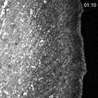
Actin dynamics in a Rac1(Q61L)-expressing PtK1 cell injected with Pak inhibitory fragment PBD/ID(H83L). Fast retrograde flow still occurs in the lamellipodium, but the width of the lamellipodium is re...
CIL:11836
NCBI Organism Classification
Potorous tridactylus
Biological Process
microtubule polymerization
Cellular Component
microtubule
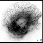
Microtubules in a constitutively active Rac1(Q61L)-expressing PtK1 cell. Microtubules are subjected to continuous retrograde flow away from the cell edge, but many plus ends undergo net growth and rem...
CIL:11837
NCBI Organism Classification
Potorous tridactylus
Biological Process
microtubule polymerization
Cellular Component
microtubule
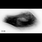
Microtubules in a dominant-negative Rac1(T17N)-expressing PtK1 cell. Microtubules do not undergo retrograde flow, and microtubules rapidly switch between growth and shortening phases with little net c...
CIL:12018
NCBI Organism Classification
Potorous tridactylus
Biological Process
attachment of spindle microtubules to kinetochore
Cellular Component
kinetochore
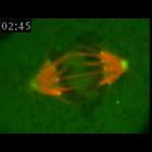
Metaphase PtK1 cell microinjected with X-rhodamine-labeled tubulin and Alexa 488-labeled CENP-F antibodies to fluorescently label kinetochore fibers (red) and kinetochores/spindle poles (green), respe...
CIL:12020
NCBI Organism Classification
Potorous tridactylus
Biological Process
attachment of spindle microtubules to kinetochore
Cellular Component
kinetochore
Mitotic PtK1 cell microinjected with X-rhodamine-labeled tubulin and Alexa 488-labeled CENP-F antibodies to fluorescently label kinetochore fibers (red) and kinetochores/spindle poles (green), respect...
CIL:11562
NCBI Organism Classification
Potorous tridactylus
Biological Process
centriole replication
Cellular Component
centriole
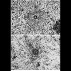
Figs. 307 & 308 from Don Fawcett's Chapter 12 (Centrioles). In interphase cells, microtubules commonly radiate from the centrosome, and in dividing cells the microtubules of the mitotic spindle conver...
CIL:12019
NCBI Organism Classification
Potorous tridactylus
Biological Process
attachment of spindle microtubules to kinetochore
Cellular Component
kinetochore
Mitotic PtK1 cell microinjected with X-rhodamine-labeled tubulin and Alexa 488-labeled CENP-F antibodies to fluorescently label kinetochore fibers (red) and kinetochores/spindle poles (green), respect...
CIL:8078
NCBI Organism Classification
Potorous tridactylus
Biological Process
attachment of spindle microtubules to kinetochore
Cellular Component
kinetochore
Metaphase PtK1 cell microinjected with X-rhodamine-labeled tubulin and Alexa 488-labeled CENP-F antibodies, to fluorescently label kinetochore fibers (red) and kinetochores/spindle poles (green), resp...
CIL:8079
NCBI Organism Classification
Potorous tridactylus
Biological Process
attachment of spindle microtubules to kinetochore
Cellular Component
kinetochore
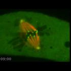
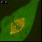
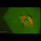
The movie starts in metaphase and shows sister kinetochore pairs aligned at the metaphase plate and oscillating between the two spindle poles as their respective kinetochore fibers lengthen and shorte...
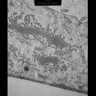
CIL:7743
NCBI Organism Classification
Potorous tridactylus tridactylus
Biological Process
protein maturation
Cellular Component
Golgi apparatus
Details of the Golgi apparatus seen in 40 nm epon sections of Ptk tissue culture cells after plunge freezing,and freeze substitution. Image recorded at 28,500x with a Philips CM10 TEM operated at 80KV
« Previous
1
2
3
Next »
Results per page:
10
20
50
100