Alternate header for print version
Contributors
Help
Submit
Search
menu
Data sets
Videos
Latest data
Center for Research in Biological Systems
Basic Science Building, Room 1000
University of California, San Diego
9500 Gilman Drive
La Jolla, CA 92093-0608, USA
Voice
: (858) 534-0276
Fax
: (858) 534-7497
Email
: dorloff@ncmir.ucsd.edu
Search Results for
scanning electron microscopy (SEM)
(381 results)
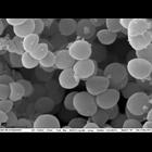
CIL:53298
NCBI Organism Classification
Prokaryote
Biological Process
none specified
Cellular Component
Cell surphace morphology
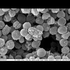
Control-treated MRSA biofilm
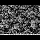
CIL:53299
NCBI Organism Classification
Prokaryote
Biological Process
none specified
Cellular Component
Cell surphace morphology
MRSA treated 3.5KX
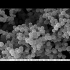
CIL:53297
NCBI Organism Classification
Prokaryote
Biological Process
none specified
Cellular Component
Cell surphace morphology
Control-treated MRSA biofilm
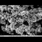
CIL:53300
NCBI Organism Classification
Prokaryote
Biological Process
none specified
Cellular Component
Cell surphace morphology
MRSA treated 3.5KX
CIL:53301
NCBI Organism Classification
Prokaryote
Biological Process
none specified
Cellular Component
Cell surphace morphology
MRSA treated 3.5KX
CIL:13091
NCBI Organism Classification
Mus musculus
Biological Process
immune system process
Cellular Component
microvillus
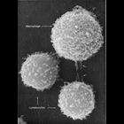
This image shows two different types of white blood cells from a mouse which are essential for mammalian immune response to protect from infection. The larger cell is a macrophage and the smaller two...
CIL:10917
NCBI Organism Classification
Rattus
Biological Process
plasma membrane organization
Cellular Component
plasma membrane
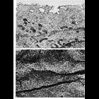
Transitional epithelial cells of the bladder. Upper panel is an electron micrograph from thin sectioned superficial cells of the uroepithelium in the rat; lower panel shows a scanning electron microg...
CIL:25356
NCBI Organism Classification
Hydra viridis
Biological Process
none specified
Cellular Component
none specified
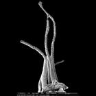
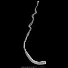
Scanning electron micrograph showing tentacles on the head of a Hydra viridis. Epithelial cells located at the base of the tentacle are in a flat, contracted state. Cell distal to the head of the H. v...
CIL:24842
NCBI Organism Classification
Hydra viridis
Biological Process
none specified
Cellular Component
none specified
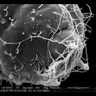
Scanning electron micrograph showing the tip of a Hydra viridis tentacle. A meshwork of triggered nematocyst cells is visible. When hooked cnidocytes on the surface of the tentacle come in contact wit...
CIL:25357
NCBI Organism Classification
Hydra viridis
Biological Process
none specified
Cellular Component
none specified
Scanning electron micrograph showing tentacles on the head of a Hydra viridis. Epithelial cells located at the base of the tentacle are in a flat, contracted state. Cell distal to the head of the H. v...
« Previous
1
2
3
4
5
6
7
8
9
...
39
Next »
Results per page:
10
20
50
100