Alternate header for print version
Contributors
Help
Submit
Search
menu
Data sets
Videos
Latest data
Center for Research in Biological Systems
Basic Science Building, Room 1000
University of California, San Diego
9500 Gilman Drive
La Jolla, CA 92093-0608, USA
Voice
: (858) 534-0276
Fax
: (858) 534-7497
Email
: dorloff@ncmir.ucsd.edu
Search Results for
scanning electron microscopy (SEM)
(381 results)
CIL:40608
NCBI Organism Classification
Mus musculus
Biological Process
none specified
Cellular Component
cell surface
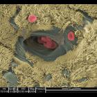
Colorized scanning electron micrograph of a mouse trachea and its red blood cells.
CIL:40613
NCBI Organism Classification
Bacillariophyta
Biological Process
frustule organization
Cellular Component
frustule
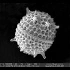
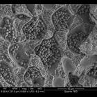
Scanning electron micrograph of a diatom frustule. The frustule is the hard and porous cell wall or external layer of diatoms.
CIL:40616
NCBI Organism Classification
Lilium
Biological Process
none specified
Cellular Component
pollen coat
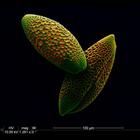
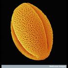
Colorized scanning electron micrograph of a Lilium pollen mounted on a carbon pad.
CIL:39021
NCBI Organism Classification
none specified
Biological Process
cell-cell adhesion
Cellular Component
cell surface
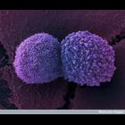
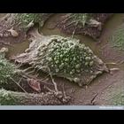
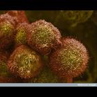
Colorized scanning electron micrograph showing two lung cancer cells. These cells were grown using cell culture techniques.
CIL:39048
NCBI Organism Classification
Paeonia
Biological Process
none specified
Cellular Component
pollen wall
Colorized scanning electron micrograph of a a grain of pollen from a peony plant. Peonies are herbaceous plants native to Asia, Southern Europe and Northern America.
CIL:39050
NCBI Organism Classification
none specified
Biological Process
none specified
Cellular Component
cell surface
Colorized scanning electron micrograph of a lung cancer cell grown in culture.
CIL:39084
NCBI Organism Classification
Lactuca sativa
Biological Process
respiratory gaseous exchange
Cellular Component
stoma
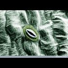
Colorized scanning electron micrograph of an open stoma on the surface of a leaf of lettuce (Lactuca sativa). A stoma is a small pore found on the underside of plant leaves and stems that allow for ga...
CIL:39110
NCBI Organism Classification
Drosophila
Biological Process
compound eye development
Cellular Component
none specified
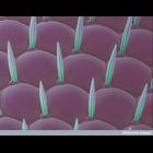
Scanning electron micrograph of the Drosophila compound eye. Close-up shows the multi-faceted cornea (pink) and the thin, hair-like structures called setae (green) which are believed to reduce glare.
CIL:39076
NCBI Organism Classification
none specified
Biological Process
cell-cell adhesion
Cellular Component
cell surface
A colorized scanning electron micrograph of pancreatic cancer cells grown in culture. See additional image at CIL: 39104.
CIL:41037
NCBI Organism Classification
Corallinales
Biological Process
none specified
Cellular Component
cell surface
Scanning electron micrograph of coralline algae. Coralline algae are characterized by a thallus that is hard because of calcareous deposits contained within the cell walls.
« Previous
1
...
3
4
5
6
7
8
9
10
...
39
Next »
Results per page:
10
20
50
100