Alternate header for print version
Contributors
Help
Submit
Search
menu
Data sets
Videos
Latest data
Center for Research in Biological Systems
Basic Science Building, Room 1000
University of California, San Diego
9500 Gilman Drive
La Jolla, CA 92093-0608, USA
Voice
: (858) 534-0276
Fax
: (858) 534-7497
Email
: dorloff@ncmir.ucsd.edu
Search Results for
vesicle membrane
(79 results)
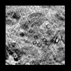
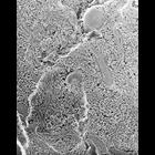
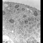
CIL:36261
NCBI Organism Classification
Vorticella convallaria
Biological Process
macronucleus organization
Cellular Component
macronucleus
Only the macronucleus contains nucleoli which is the site of ribosomal RNA synthesis and ribosome subunit assembly. This EM also contains the distal end of the food vacuole forming cytopharynx which c...
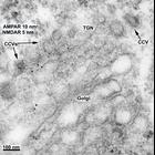
CIL:11923
NCBI Organism Classification
Rattus
Biological Process
none specified
Cellular Component
clathrin coat of trans-Golgi network vesicle
NMDA receptors (NMDARs) can exit the Golgi/TGN via clathrin-coated vesicles. Double-labeling using rabbit polyclonal antibodies to AMPA receptors (AMPARs) (combination of 3 antibodies: GluR1, GluR2, G...
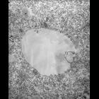
CIL:41362
NCBI Organism Classification
Mus musculus
Biological Process
cytoplasm organization
Cellular Component
mitochondrion
The image shows a slice through a 3D tomographic reconstruction of a mouse adenocarcinoma cell based on x-ray microscopy. Live cells were cryofixed by plunge freezing in liquid ethane and the vitrifi...
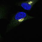
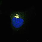
CIL:13564
NCBI Organism Classification
Homo sapiens
Biological Process
retrograde vesicle-mediated transport, Golgi to ER
Cellular Component
Golgi cisterna membrane
COP-I beta1 (green) staining at the Golgi (GM130, a cis-Golgi marker) (red) in unstimulated HeLa cells. Helix Pomatia Lectin (HPL) (gray) staining is also shown. HPL binds various glycans but the Tn a...
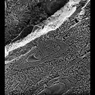
CIL:39720
NCBI Organism Classification
Tetrahymena pyriformis
Biological Process
phagocytosis, engulfment
Cellular Component
phagocytic vesicle
The nascent digestive vacuole forms as the membrane of flattened vesicles fuse with the single membrane of the cytopharynx between the lamellae (2 microtubules forming a lamella) that are connected al...
CIL:12615
NCBI Organism Classification
Paramecium multimicronucleatum
Biological Process
cortical cytoskeleton organization
Cellular Component
cell cortex
High resolution micrograph following trifluoperizine (TFP) treatment, which apparently does not inhibit endocytosis but does inhibit movement of vesicles away from the parasomal sacs, coated and preen...
CIL:21606
NCBI Organism Classification
Paramecium multimicronucleatum
Biological Process
endosome organization
Cellular Component
cytoplasm
Quick-freeze deep-etch rotary-shadowed replica of an early endosome with two clathrin-coated evaginations. Presumably these coated pits contain receptors for cargo that will be pinched off from the ea...
CIL:12634
NCBI Organism Classification
Paramecium multimicronucleatum
Biological Process
endosome organization
Cellular Component
coated pit
High resolution image of two parasomal sacs that are oriented perpendicular to the basal bodies. The top parasomal sac has a clathrin cage around it's coated pit area. The P-face of the plasma membran...
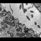
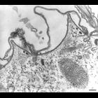
CIL:17447
NCBI Organism Classification
Paramecium multimicronucleatum
Biological Process
clathrin coating of Golgi vesicle, plasma membrane to endosome targeting
Cellular Component
cell cortex
Cell sectioned perpendicular to the surface. The parasomal sac ends internally as a coated pit, coated with clathrin. Where the coated pit has pinched off a clathrin-coated vesicle is formed. When the...
CIL:13565
NCBI Organism Classification
Homo sapiens
Biological Process
retrograde vesicle-mediated transport, Golgi to ER
Cellular Component
Golgi cisterna membrane
COP-I gamma1 (green) staining at the Golgi (GM130, a cis-Golgi marker) (red) in unstimulated HeLa cells. Helix Pomatia Lectin (HPL) (gray) staining is also shown. HPL binds various glycans but the Tn ...
« Previous
1
2
3
4
5
6
7
8
Next »
Results per page:
10
20
50
100

")