Alternate header for print version
Contributors
Help
Submit
Search
menu
Data sets
Videos
Latest data
Center for Research in Biological Systems
Basic Science Building, Room 1000
University of California, San Diego
9500 Gilman Drive
La Jolla, CA 92093-0608, USA
Voice
: (858) 534-0276
Fax
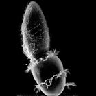
: (858) 534-7497
Email
: dorloff@ncmir.ucsd.edu
Search Results for
plasma membrane part
(338 results)
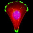
CIL:12655
NCBI Organism Classification
Homo sapiens
Biological Process
substrate adhesion-dependent cell spreading
Cellular Component
focal adhesion
Human primary keratinocyte spreading on an asymmetric "arc"-shaped collagen I micropattern. The cells are stained for the focal adhesion protein vinculin (green, Alexa488), F-Actin (red, TRITC phallo...
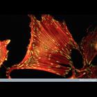
CIL:38985
NCBI Organism Classification
Rattus
Biological Process
cell adhesion
Cellular Component
stress fiber
Fluorescent micrograph of rat embryo fibroblast cell growing in serum stained to reveal actin stress fibres (red) and vinculin (component of focal adhesions) in green/yellow.
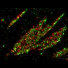
CIL:38651
NCBI Organism Classification
Homo sapiens
Biological Process
molecular organization
Cellular Component
focal adhesion
This photoactivation localization microscopy (PALM) image of tdEos-vinculin (red) and Dronpa-paxillin (green) illustrates that vinculin and paxillin are segregated into interlocking microdomains withi...
CIL:11237
NCBI Organism Classification
Macaca mulatta
Biological Process
cell communication
Cellular Component
gap junction
Representative examples of gap junctions from vertebrates (ciliary epithelium from the eye of a Macaca mulatta, upper) and invertebrates (inverted gap junctions from cells of the mid-gut of the horses...
CIL:17891
NCBI Organism Classification
Didinium nasutum
Biological Process
phagocytosis
Cellular Component
oral apparatus
The ingestion of Paramecium by Didinium. This micrograph also clearly shows the five short rows of clavate cilia (ciliary stubs without central microtubules) that reside just below the anterior and po...
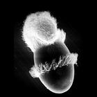
CIL:19535
NCBI Organism Classification
Didinium nasutum
Biological Process
phagocytosis
Cellular Component
oral apparatus
Didinium captures Paramecium. Also showing metachronous waves of cilia in the two characteristic ciliary girdles of Didinium nasutum. This micrograph was taken in 1968 by G. Antipa on a Cambridge Mar...
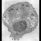
CIL:36228
NCBI Organism Classification
Tetrahymena pyriformis
Biological Process
digestive system process
Cellular Component
cytostome
Tetrahymena cells that are not forming a digestive vacuole often have a tube-like extension of the cytopharyngeal membrane attached to the deep fiber that extends several microns into the cytoplasm. T...
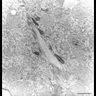
CIL:9826
NCBI Organism Classification
Opercularia [NCBITaxon:168247]
Biological Process
macronucleus organization
Cellular Component
macronucleus
This figure shows the connection between the peristome and the food vacuole forming zone where the alveoli end. The cytostome is the opening that connects these two zones. TEM taken on 6/13/69 by R. ...
CIL:38975
NCBI Organism Classification
Homo sapiens
Biological Process
cell adhesion to artificial surface
Cellular Component
cell surface
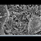
Scanning electron micrograph of two human bone-forming cells (osteoblasts) crawling over crystals of the ceramic material,. monetite (CaHPO4). Monetite crystals are electrochemically deposited onto ti...
CIL:42801
NCBI Organism Classification
none specified
Biological Process
none specified
Cellular Component
cell surface
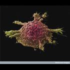
Colorized scanning electron micrograph of a cell cultured lung cancer cell. This image is part of an image group, CIL 42801-42803, showing several colorized scanning electron micrographs of cell cult...
« Previous
1
...
5
6
7
8
9
10
11
12
...
34
Next »
Results per page:
10
20
50
100