Alternate header for print version
Contributors
Help
Submit
Search
menu
Data sets
Videos
Latest data
Center for Research in Biological Systems
Basic Science Building, Room 1000
University of California, San Diego
9500 Gilman Drive
La Jolla, CA 92093-0608, USA
Voice
: (858) 534-0276
Fax
: (858) 534-7497
Email
: dorloff@ncmir.ucsd.edu
Search Results for
plasma membrane part
(338 results)
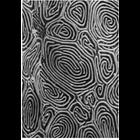
CIL:11110
NCBI Organism Classification
Parophrys vetulus
Biological Process
plasma membrane organization
Cellular Component
cell surface
of 'The Cell, 2nd Ed.' by Don W. Fawcett M.D. Scanning electron micrograph of the epidermal surface of the flatfish sole Parophrys detulus shows a labyrinth of surface ridges. Image from Fahrenbach,...
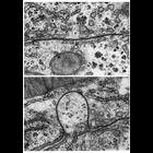
CIL:11235
NCBI Organism Classification
none specified
Biological Process
cell communication
Cellular Component
gap junction
Upper: a gap junction on the boundary between two hepatic cells shows a typical narrow and straight interface. Lower: in some preparations, gap junctions appear curved, and project into one of the ce...
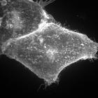
CIL:9098
NCBI Organism Classification
Homo sapiens
Biological Process
cellular membrane organization
Cellular Component
anchored to plasma membrane
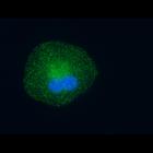
Hela cells transiently transfected with EGFP-GL-GPI show the organization of the membrane thru the distribution of GPI anchored protein. This image is the maximum z projection image that accompanies...
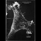
CIL:11129
NCBI Organism Classification
Mus musculus
Biological Process
cell motility
Cellular Component
lamellipodium membrane
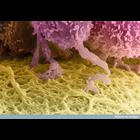
Scanning electron micrograph of a 3T3 cell growing in culture. Complexity and diversity of structure across the cell surface is apparent in the this image that s courtesy of Susan Brown. Figure 48 fr...
CIL:38957
NCBI Organism Classification
Homo sapiens
Biological Process
receiving nourishment
Cellular Component
zona pellucida
A colorized scanning electron micrograph of a human egg, which is the huge cell colored yellow at the bottom of this image. The follicle cells that surround it (top) send out long projections that pen...
CIL:9097
NCBI Organism Classification
Homo sapiens
Biological Process
cellular membrane organization
Cellular Component
anchored to plasma membrane
Hela cells transiently transfected with EGFP-GL-GPI show the organization of the membrane thru the distribution of GPI anchored protein. This image is the z stack that accompanies the related maximu...
CIL:11115
NCBI Organism Classification
Entosphenus tridentatus
Biological Process
plasma membrane organization
Cellular Component
cell surface
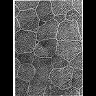
Scanning electron micrograph of the epidermal surface of lamprey larvae. A row of microvilli outline the polygonal borders between cells, while short microvilli cover the external surface in a reticu...
CIL:10103
NCBI Organism Classification
Homo sapiens
Biological Process
none specified
Cellular Component
AP-2 adaptor complex
Cultured retinal pigment epithelial cells immunofluorescently labeled for adaptor protein-2 (AP2) (green) and nucleus (blue). The cells were fixed in 2% PFA and 0.5% Triton X-100 for 2 minutes follow...
CIL:11199
NCBI Organism Classification
Rattus
Biological Process
cell-cell adhesion
Cellular Component
zonula adherens
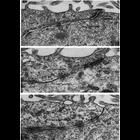
These examples of ependymal epithelium from the rat brain represent epithelia that lack zonulae occludentes. Further, there appears to be no consistent order to the junctional specializations that are...
CIL:11201
NCBI Organism Classification
Siphonaptera
Biological Process
cell-cell junction organization
Cellular Component
septate junction
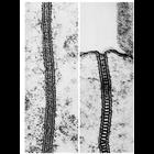
Epithelia of invertebrates contain zona continua and septate junctions not found in mammals. Left, zonula continua from gut epithelium of a flea; right, septate junction from the midgut epithelium of...
« Previous
1
2
3
4
5
6
7
8
9
...
34
Next »
Results per page:
10
20
50
100