Alternate header for print version
Contributors
Help
Submit
Search
menu
Data sets
Videos
Latest data
Center for Research in Biological Systems
Basic Science Building, Room 1000
University of California, San Diego
9500 Gilman Drive
La Jolla, CA 92093-0608, USA
Voice
: (858) 534-0276
Fax
: (858) 534-7497
Email
: dorloff@ncmir.ucsd.edu
Search Results for
microtubule organizing center part
(91 results)
CIL:35425
NCBI Organism Classification
Mus musculus
Biological Process
meiotic spindle organization
Cellular Component
microtubule
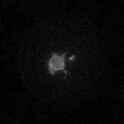
Time-lapse microscopy of histone-RFP expressing mouse oocytes injected with siCo-5h, a control siRNA. Images were taken every 20 minutes. Times after the resumption of meiotic maturation and germinal ...
CIL:35424
NCBI Organism Classification
Mus musculus
Biological Process
meiotic spindle organization
Cellular Component
microtubule
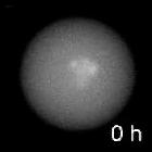
Time-lapse microscopy of b-Tubulin GFP expressing mouse oocytes injected with siTPX2-5h, a doublestranded siRNA against TPX2 (targeting protein for Xenopus kinesin-like protein, xklp2). Images were ta...
CIL:35428
NCBI Organism Classification
Mus musculus
Biological Process
meiotic spindle organization
Cellular Component
microtubule
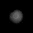
Time-lapse microscopy of mouse oocytes expressing YFP-TPX2DN, a truncated version of TPX2 (targeting protein for Xenopus kinesin-like protein, xklp2) which lacks the 118 first amino acids and injected...
CIL:35430
NCBI Organism Classification
Mus musculus
Biological Process
meiotic spindle organization
Cellular Component
microtubule
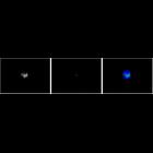
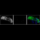
This 3 panel image displays meiosis in a mouse oocyte triple labelled for microtubules (panel 1 and green), phosphorylated-Transforming Acidic Coiled-Coil protein (TACC3; panel 2 and red), and nuclei...
CIL:35432
NCBI Organism Classification
Mus musculus
Biological Process
meiotic spindle organization
Cellular Component
microtubule
This 3 panel image displays meiosis in a mouse oocyte triple labelled for microtubules (panel 1 and green), phosphorylated-Transforming Acidic Coiled-Coil protein (TACC3; panel 2 and red), and nuclei...
CIL:11556
NCBI Organism Classification
Gallus gallus
Biological Process
spermatogonial cell division
Cellular Component
pericentriolar material
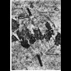
Figure 302 from Chapter 12 (Centrioles) by Don Fawcett. The centrioles replicate early in cell division and take up positions at either pole of the division figure. Concurrently with the condensation ...
CIL:11562
NCBI Organism Classification
Potorous tridactylus
Biological Process
centriole replication
Cellular Component
centriole
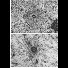
Figs. 307 & 308 from Don Fawcett's Chapter 12 (Centrioles). In interphase cells, microtubules commonly radiate from the centrosome, and in dividing cells the microtubules of the mitotic spindle conver...
CIL:11564
NCBI Organism Classification
Cavia porcellus
Biological Process
centriole satellites
Cellular Component
centriole
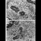
Figs. 309 & 310 from Don Fawcett's Chapter 12 (Centrioles). The plane of a thin section only rarely happens to coincide with the long axis of both members of a pair of centrioles. It is more common fo...
CIL:37204
NCBI Organism Classification
Cavia porcellus
Biological Process
none specified
Cellular Component
atrial granules
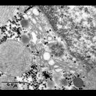
Transmission electron micrograph of section of cardiac muscle from guinea pig right atrium. A portion of nucleus is seen at upper right, with a centriole close to the nuclear envelope. Also seen in ...
CIL:7723
NCBI Organism Classification
Cricetulus griseus
Biological Process
mitosis
Cellular Component
centriole
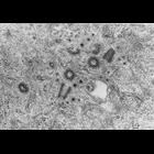
Four centrioles imaged in one electron micrograph of a thin section, cut from a Chinese hamster fibroblast grown in tissue culture. The two centrioles that appear circular are the 'mother' centrioles,...
« Previous
1
2
3
4
5
6
7
8
9
10
Next »
Results per page:
10
20
50
100