Alternate header for print version
Contributors
Help
Submit
Search
menu
Data sets
Videos
Latest data
Center for Research in Biological Systems
Basic Science Building, Room 1000
University of California, San Diego
9500 Gilman Drive
La Jolla, CA 92093-0608, USA
Voice
: (858) 534-0276
Fax
: (858) 534-7497
Email
: dorloff@ncmir.ucsd.edu
Search Results for
lysosome
(152 results)
CIL:46703
NCBI Organism Classification
Homo sapiens
Biological Process
none specified
Cellular Component
peroxisome
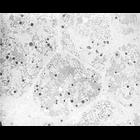
Low magnification electron micrograph showing peroxisomes in several parenchymal cells in human liver, contrasted by catalase activity with DAB. A few organelles show a heterogeneous content: they are...
CIL:46706
NCBI Organism Classification
Homo sapiens
Biological Process
regulation of phosphatase activity
Cellular Component
peroxisome
Normal morphology of two peroxisomes (marked P) in a human liver parenchymal cell. They are contiguous with a narrow endoplasmic reticulum cisterna; this relationship is frequent. The image also shows...
CIL:10839
NCBI Organism Classification
Phodopus
Biological Process
autophagy
Cellular Component
lysosome
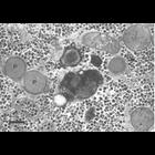
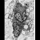
Figure 263 from Chapter 8 (Lysosomes) of 'The Cell, 2nd Ed.' by Don W. Fawcett M.D. Portion of a cell from hamster suprarenal cortex with clustered lysosomes near the Golgi complex. A PDF copy of th...
CIL:11378
NCBI Organism Classification
Leporidae
Biological Process
post-translational protein modification
Cellular Component
Golgi apparatus
Figure 210 from Chapter 6 (Golgi Apparatus) of 'The Cell, 2nd Ed.' by Don W. Fawcett M.D. Supranuclear region of a cell from rabbit epididymis. Epithelial cells of the epididymis have a large Golgi a...
CIL:10845
NCBI Organism Classification
Leporidae
Biological Process
autophagy
Cellular Component
lysosome
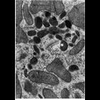
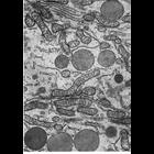
Figure 271 from Chapter 8 (Lysosomes) of 'The Cell, 2nd Ed.' by Don W. Fawcett M.D. Apical region of epithelial cells from the proximal portion of the cauda epididymis of rabbit shows both large, den...
CIL:11133
NCBI Organism Classification
Didelphimorphia
Biological Process
pinocytosis
Cellular Component
lysosome
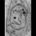
A thin section through a macrophage from interstitial tissue of the testes of the opossum. These cells are characterized by extensive folding of the cell surface, some of which appear to have captured...
CIL:10843
NCBI Organism Classification
Spermophilus citellus
Biological Process
autophagy
Cellular Component
lysosome
Figure 270 from Chapter 8 (Lysosomes) of 'The Cell, 2nd Ed.' by Don W. Fawcett M.D. Cytoplasm from a cell from a ductus efferens of the ground squirrel Citellus lateralis with spherical membrane-encl...
CIL:35999
NCBI Organism Classification
Gerbillinae
Biological Process
secretory granule organization
Cellular Component
secretory granule
Figures 379 (upper) and 380 (lower) from Chapter 15 (Cytoplasmic Inclusions) of 'The Cell, 2nd Ed.' by Don W. Fawcett M.D. Freeze substitution preparations of gerbil parotid gland show condensing vac...
CIL:40430
NCBI Organism Classification
Saccharomyces cerevisiae
Biological Process
organelle organization
Cellular Component
autophagic vacuole
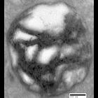
Transmission electron micrograph of an autophago-lysosome from a sertraline-treated prototrophic wildtype cell. (40,000X). This is part of an image group highlighting multilamellar structure, CIL: 404...
CIL:37335
NCBI Organism Classification
none specified
Biological Process
cell migration
Cellular Component
nucleus
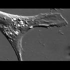
A more fully differentiated epidermal cell. The nucleus and nucleoli are on the left. The video shows the intracellular movements of lysosomes and mitochondria. The lysosomes are the small refractile ...
« Previous
1
2
3
4
5
6
7
8
9
...
16
Next »
Results per page:
10
20
50
100