Alternate header for print version
Contributors
Help
Submit
Search
menu
Data sets
Videos
Latest data
Center for Research in Biological Systems
Basic Science Building, Room 1000
University of California, San Diego
9500 Gilman Drive
La Jolla, CA 92093-0608, USA
Voice
: (858) 534-0276
Fax
: (858) 534-7497
Email
: dorloff@ncmir.ucsd.edu
Search Results for
lamellipodium
(146 results)
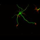
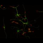
CIL:10224
NCBI Organism Classification
Rattus
Biological Process
developmental process
Cellular Component
cytoskeleton
This multi-layer image shows the spatial relationship between filamentous actin (red) and microtubule array (green) in cultured hippocampal neurons, grown for 3 days in vitro. Actin staining (with rh...
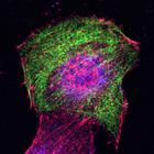
CIL:26526
NCBI Organism Classification
Mus musculus
Biological Process
regulation of cell migration
Cellular Component
lamellipodium
To study the molecular mechanism by which nonmuscle myosin II (MII) regulates protrusion and adhesion dynamics in migrating cells, NIH3T3 cells were co-stained for βPIX (green) and myosin IIA (red). ...
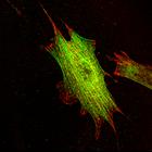
CIL:26528
NCBI Organism Classification
Mus musculus
Biological Process
regulation of cell migration
Cellular Component
lamellipodium
To study the molecular mechanism by which nonmuscle myosin II (MII) regulates protrusion and adhesion dynamics in migrating cells, NIH3T3 cells were transfected with myc-tagged myosin light chain (MLC...
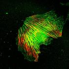
CIL:26549
NCBI Organism Classification
Mus musculus
Biological Process
regulation of cell migration
Cellular Component
lamellipodium
To study the molecular mechanism by which nonmuscle myosin II (MII) regulates protrusion and adhesion dynamics in migrating cells, Swiss3T3 cells were co-stained for βPIX (green) and myosin IIA (red)...
CIL:26557
NCBI Organism Classification
Mus musculus
Biological Process
regulation of cell migration
Cellular Component
lamellipodium
To study the molecular mechanism by which nonmuscle myosin II (MII) regulates protrusion and adhesion dynamics in migrating cells, NIH3T3 cells were treated with DMSO for 15 min. and stained for βPIX...
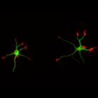
CIL:8785
NCBI Organism Classification
Rattus
Biological Process
developmental process
Cellular Component
cytoskeleton
This color combined image shows the spatial relationship between filamentous actin (red) and microtubule array (green) in cultured hippocampal neurons, grown for 1 day in vitro. Actin staining (with ...
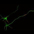
CIL:8780
NCBI Organism Classification
Rattus
Biological Process
developmental process
Cellular Component
cytoskeleton
This color combined image shows the spatial relationship between filamentous actin (red) and microtubule array (green) in cultured hippocampal neurons, grown for 1 day in vitro. Actin staining (with ...
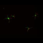
CIL:10094
NCBI Organism Classification
Rattus
Biological Process
developmental process
Cellular Component
cytoskeleton
This multi-layer image shows the spatial relationship between filamentous actin (red) and microtubule array (green) in cultured hippocampal neurons, grown for 1 day in vitro. Actin staining (with rho...
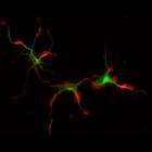
CIL:10110
NCBI Organism Classification
Rattus
Biological Process
developmental process
Cellular Component
cytoskeleton
This multi-layer image shows the spatial relationship between filamentous actin (red) and microtubule array (green) in cultured hippocampal neurons, grown for 3 days in vitro. Actin staining (with rh...
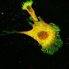
CIL:10223
NCBI Organism Classification
Rattus
Biological Process
developmental process
Cellular Component
cytoskeleton
This multi-layer image shows the spatial relationship between filamentous actin (red) and microtubule array (green) in cultured hippocampal neurons, grown for 3 days in vitro. Actin staining (with rh...
« Previous
1
...
3
4
5
6
7
8
9
10
...
15
Next »
Results per page:
10
20
50
100