Alternate header for print version
Contributors
Help
Submit
Search
menu
Data sets
Videos
Latest data
Center for Research in Biological Systems
Basic Science Building, Room 1000
University of California, San Diego
9500 Gilman Drive
La Jolla, CA 92093-0608, USA
Voice
: (858) 534-0276
Fax
: (858) 534-7497
Email
: dorloff@ncmir.ucsd.edu
Search Results for
extracellular region
(191 results)
CIL:25687
NCBI Organism Classification
Trichodina
Biological Process
response to symbiont
Cellular Component
symbiont-containing vacuole
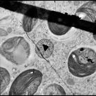
Cytoplasm of a green Trichodina that lives on the surface of a freshwater snail collected in central Illinois. In this view the abundant endosymbiotic zoochlorellae and a single endosymbiotic bacteriu...
CIL:11529
NCBI Organism Classification
Pongo pygmaeus
Biological Process
pigmentation
Cellular Component
melanosome
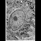
Figure 297 from Chapter 11 (Melanin Pigment) of 'The Cell, 2nd Ed.' by Don W. Fawcett M.D. Epidermal melanocyte from facial skin of an orangutan illustrates the interstitial location of a melanocyte ...
CIL:39796
NCBI Organism Classification
Tetrahymena pyriformis
Biological Process
microtubule basal body duplication
Cellular Component
microtubule basal body
Basal body formation occurs most prominently in the equatorial region of the predivision cell. A new basal body begins to develop perpendicular to the mature basal body at its proximal anterior surfac...
CIL:19117
NCBI Organism Classification
Spiroplasma kunkelii
Biological Process
pathogenesis
Cellular Component
basal lamina
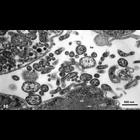
Electron micrograph of corn stunt spiroplasma (CSS) in the wall of the filter chamber (part of the alimentary canal) and in the hemolymph (he) of a vector leafhopper D. gelbus. Note the mainly pleiomo...
CIL:27229
NCBI Organism Classification
Fundulus heteroclitus
Biological Process
extracellular matrix organization
Cellular Component
fibrillar collagen
Collagen fibrils and mineralization in the external layer of Fundulus heteroclitus scale. The external layer is the second of three layers closest to the dermis of an elasmoid. It is composed of thin ...
CIL:10937
NCBI Organism Classification
none specified
Biological Process
extracellular structure organization
Cellular Component
plasma membrane
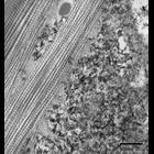
These two micrographs show the outer cell surface of the epithelial cell border in intestine (upper) and stomach (lower) tissue. This surface, called the glycocalyx, is composed of negatively charged...
CIL:10929
NCBI Organism Classification
Felis catus
Biological Process
extracellular structure organization
Cellular Component
plasma membrane
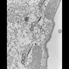
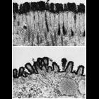
This electron micrograph shows a region of the brush border of the cat intestine. Tufted and branched polysaccharide filaments, each a few nanometers thick, extend from the microvilli to make up the ...
CIL:10932
NCBI Organism Classification
Felis catus
Biological Process
extracellular structure organization
Cellular Component
plasma membrane
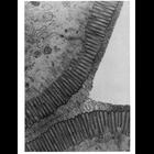
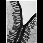
This electron micrograph highlights a darkly-stained glycocalyx rim of the brush border of the intestinal epithelium of the cat, stained en bloc with colloidal thorium. The glycocalyx is composed of ...
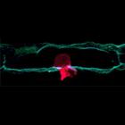
CIL:134
NCBI Organism Classification
Caenorhabditis elegans
Biological Process
cell migration
Cellular Component
basement membrane
The image shows an anchor cell (red) in the developing female reproductive system of C. elegans pushing through the basement membrane (green) that surrounds it. This type of invasion is also often see...
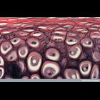
CIL:39091
NCBI Organism Classification
none specified
Biological Process
cartilage morphogenesis
Cellular Component
fibrillar collagen
Light micrograph of elastic cartilage of the ear. This type of cartilage is made from a protein called elastin and is formed from a network of elastic fibers and collagen. The fibers form bundles whic...
« Previous
1
...
3
4
5
6
7
8
9
10
...
20
Next »
Results per page:
10
20
50
100