Alternate header for print version
Contributors
Help
Submit
Search
menu
Data sets
Videos
Latest data
Center for Research in Biological Systems
Basic Science Building, Room 1000
University of California, San Diego
9500 Gilman Drive
La Jolla, CA 92093-0608, USA
Voice
: (858) 534-0276
Fax
: (858) 534-7497
Email
: dorloff@ncmir.ucsd.edu
Search Results for
cytoplasmic vesicle
(448 results)
CIL:36749
NCBI Organism Classification
Paramecium multimicronucleatum
Biological Process
digestive system process
Cellular Component
phagolysosome
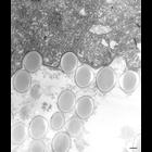
A DV-III contains a thick continuous glycocalyx (polysaccharide coat) on the luminal side of its membrane as well as paracrystalline material, both provided by the fused lysosomes. The latex beads ind...
CIL:10244
NCBI Organism Classification
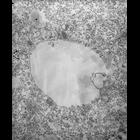
Halteria grandinella
Biological Process
macronucleus organization
Cellular Component
macronucleus
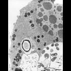
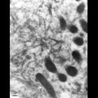
This image of Halteria was fixed in cacodylate-buffered fixative and stained. The round, dark bodies, possibly pigment granules, were transparent in other preparations are electron-opaque following th...
CIL:12614
NCBI Organism Classification
Paramecium multimicronucleatum
Biological Process
cortical cytoskeleton organization
Cellular Component
cell cortex
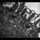
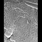
A high resolution image of a cell actively undergoing endocytosis that develops an extensive layer of vesicles and cisternae composed for the most part of smooth undecorated membrane. The uncoated cis...
CIL:13564
NCBI Organism Classification
Homo sapiens
Biological Process
retrograde vesicle-mediated transport, Golgi to ER
Cellular Component
Golgi cisterna membrane
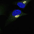
COP-I beta1 (green) staining at the Golgi (GM130, a cis-Golgi marker) (red) in unstimulated HeLa cells. Helix Pomatia Lectin (HPL) (gray) staining is also shown. HPL binds various glycans but the Tn a...
CIL:21697
NCBI Organism Classification
Paramecium multimicronucleatum
Biological Process
contractile vacuole organization
Cellular Component
contractile vacuole
Quick-freeze deep-etch image of a fracture through the radial canal of the contractile vacuole. This technique presents a picture of a confusing array of non-etchable material surrounding a collecting...
CIL:36233
NCBI Organism Classification
Tetrahymena pyriformis
Biological Process
contractile vacuole organization
Cellular Component
contractile vacuole pore
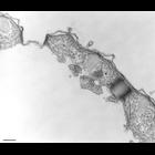
The two CV pores of a cell. An impression of the helically wound microtubules are detectable in the right pore. TEM taken on 4/21/78 by R. Allen with Hitachi HU11A operating at 60kV. Neg. 21,750X. Bar...
CIL:39794
NCBI Organism Classification
Tetrahymena pyriformis
Biological Process
contractile vacuole organization
Cellular Component
contractile vacuole pore
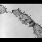
The two CV pores of a cell. An impression of the helically wound microtubules are detectable in the right pore. TEM taken on 4/21/78 by R. Allen with Hitachi HU11A operating at 60kV. Neg. 21,750X. The...
CIL:27151
NCBI Organism Classification
Paramecium multimicronucleatum
Biological Process
contractile vacuole organization
Cellular Component
contractile vacuole
An electron micrograph of a thin frozen section treated with a clone of the mAb (D40-2) used to show the A4 antigen on decorated tubules. The immunogold lies along the outer margin of these tubules fo...
CIL:9700
NCBI Organism Classification
Coleps hirtus
Biological Process
water transport
Cellular Component
contractile vacuole
Some tubules near a contractile vacuole which appear to be decorated with pegs similar to the spongiome of the CV of Paramecium. In Paramecium these pegs are known to be V-type proton pumps (V-ATPases...
CIL:39720
NCBI Organism Classification
Tetrahymena pyriformis
Biological Process
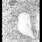
phagocytosis, engulfment
Cellular Component
phagocytic vesicle
The nascent digestive vacuole forms as the membrane of flattened vesicles fuse with the single membrane of the cytopharynx between the lamellae (2 microtubules forming a lamella) that are connected al...
« Previous
1
2
3
4
5
6
7
8
9
...
45
Next »
Results per page:
10
20
50
100