Alternate header for print version
Contributors
Help
Submit
Search
menu
Data sets
Videos
Latest data
Center for Research in Biological Systems
Basic Science Building, Room 1000
University of California, San Diego
9500 Gilman Drive
La Jolla, CA 92093-0608, USA
Voice
: (858) 534-0276
Fax
: (858) 534-7497
Email
: dorloff@ncmir.ucsd.edu
Search Results for
cell surface
(1377 results)
CIL:11105
NCBI Organism Classification
Mus musculus
Biological Process
plasma membrane organization
Cellular Component
cell surface
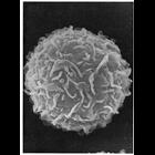
Scanning electron micrograph of the surface of a peritoneal mast cell from mouse shows undulating folds, and few microvilli. The membrane folds could be misinterpreted for microvilli if analyzed in t...
CIL:11111
NCBI Organism Classification
Paracheirodon innesi
Biological Process
cell projection organization
Cellular Component
cell surface
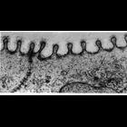
Electron micrograph showing fingerprint like ridges called microplicae on the surface of fish skin epidermis from the neon tetra. These ridges offer mechanical reinforcement against trauma and help ho...
CIL:11129
NCBI Organism Classification
Mus musculus
Biological Process
cell motility
Cellular Component
lamellipodium membrane
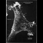
Scanning electron micrograph of a 3T3 cell growing in culture. Complexity and diversity of structure across the cell surface is apparent in the this image that s courtesy of Susan Brown. Figure 48 fr...
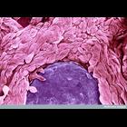
CIL:38809
NCBI Organism Classification
Homo sapiens
Biological Process
blood coagulation, fibrin clot formation
Cellular Component
extracellular matrix part
Scanning electron micrograph of a blood clot on the underside of a sticking plaster that has been used to treat a razor blade cut. Red blood cells (shown in red) and thin fibres of the protein fibrin ...
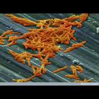
CIL:38815
NCBI Organism Classification
Clostridium difficile
Biological Process
none specified
Cellular Component
cell surface
A colour-enhanced scanning electron micrograph image showing a cluster of Clostridium difficile on a surface. Clostridium difficile is a species of Gram-positive bacteria that causes severe diarrhea a...
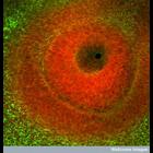
CIL:38980
NCBI Organism Classification
Gallus gallus
Biological Process
eye development
Cellular Component
cell surface
Confocal image of the developing eye of a chick embryo. The dark region in the centre is the where the surface layer has invaginated to form the lens vesicle. The tissue surrounding this space is stai...
CIL:39248
NCBI Organism Classification
Didinium nasutum
Biological Process
phagocytosis
Cellular Component
oral apparatus
Didinium captures Paramecium. Paramecium is almost completely ingested and the proboscis is beginning to reform. This micrograph was taken in 1968 by G. Antipa on a Cambridge Mark IIA operating at 20k...
CIL:39087
NCBI Organism Classification
Homo sapiens
Biological Process
none specified
Cellular Component
cell surface
Colorized scanning electron micrograph showing psoriasis (an inflammatory skin disorder) of the tongue. This condition on the tongue causes rough, scaley and bumping texture to the surface.
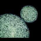
CIL:40972
NCBI Organism Classification
Nostoc commune
Biological Process
none specified
Cellular Component
cell surface
Darkfield image of spherical colonies of Nostoc commune, a bluegreen alga. Nostoc commune is a terrestrial species of cyanobacteria that in 1988 was found to harbor a previously unidentified UV-A/B ab...
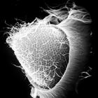
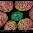
CIL:40588
NCBI Organism Classification
Porifera
Biological Process
structural support
Cellular Component
cell surface
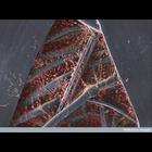
This colorized scanning electron micrograph shows uncoated sponge spicules from a South African sponge. Spicules provide structural support and deter predators. The image was taken with the VcD (back...
« Previous
1
...
6
7
8
9
10
11
12
13
...
138
Next »
Results per page:
10
20
50
100