Alternate header for print version
Contributors
Help
Submit
Search
menu
Data sets
Videos
Latest data
Center for Research in Biological Systems
Basic Science Building, Room 1000
University of California, San Diego
9500 Gilman Drive
La Jolla, CA 92093-0608, USA
Voice
: (858) 534-0276
Fax
: (858) 534-7497
Email
: dorloff@ncmir.ucsd.edu
Search Results for
cell surface
(1377 results)
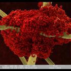
CIL:39056
NCBI Organism Classification
Mus musculus
Biological Process
bone morphogenesis
Cellular Component
cell surface
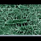
This colorized scanning electron micrograph shows the structure of osteocyte cells in the cortex of a mouse tibia bone. Osteocytes are the cells that form new bone. Imbedding the bone in resin, which ...
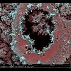
CIL:38927
NCBI Organism Classification
Rhizopus
Biological Process
formation of fruiting body
Cellular Component
cell surface
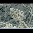
Scanning electron micrograph of the the fruiting body of bread mould. As the mould grows, it propagates itself by forming spores that are released into the environment and grow into new mould colonies...
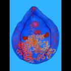
CIL:39249
NCBI Organism Classification
Didinium nasutum
Biological Process
phagocytosis
Cellular Component
oral apparatus
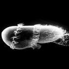
Didinium captures Paramecium. At midingestion and showing metachronous waves of cilia within the two characteristic ciliary girdles of Didinium nasutum. This micrograph was taken in 1968 by G. Antipa ...
CIL:40593
NCBI Organism Classification
Staphylococcus aureus
Biological Process
antibiotic resistance
Cellular Component
cell surface
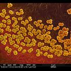
Colorized scanning electron micrograph of methicillin resistant staphylococcus aureus (MRSA) bacteria on the surface of a wound dressing.
CIL:40600
NCBI Organism Classification
none specified
Biological Process
blood coagulation
Cellular Component
cell surface
Colorized scanning electron micrograph of partially dried red blood cells clotted on the cotton fibres of a gauze wound dressing. Image was collected at low vacuum to avoid charging of cotton fibres
CIL:40618
NCBI Organism Classification
none specified
Biological Process
biofilm formation
Cellular Component
cell surface
Colorized scanning electron micrograph of a biofilm on carbon steel after immersion in sea water for 14 days. A biofilm is an aggregate of microorganisms that adhere to each other on a surface and ar...
CIL:41736
NCBI Organism Classification
Bacillus subtilis
Biological Process
biofilm organization
Cellular Component
cell surface
A confocal micrograph showing Bacillus subtilis, a Gram-positive, rod-shaped bacterium that is commonly found in soil. Distinct lineages of bacteria expressing different fluorescent proteins were init...
CIL:41839
NCBI Organism Classification
Prosthogonimus macrorchis
Biological Process
parasite infection
Cellular Component
cell surface
Prosthogonimus macrorchis, a flatworm poultry parasite, imaged with Rheinberg illumination. Honorable Mention, 2010 Olympus BioScapes Digital Imaging Competition®. Prosthogonimus macrorchis has a com...
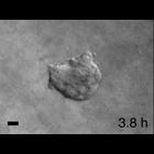
CIL:42163
NCBI Organism Classification
Homo sapiens
Biological Process
dissemination
Cellular Component
cell surface
Time-lapse movie of a human tumor fragment grown in Matrigel that was freed and reembedded in collagen I. Images collected every 15 minutes and bar is 50 microns. CIL 42162 - CIL 42165 are related m...
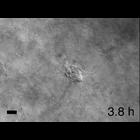
CIL:42165
NCBI Organism Classification
Homo sapiens
Biological Process
dissemination
Cellular Component
cell surface
Time-lapse movie of a human tumor fragment grown in colllagen I that was freed and reembedded in collagen I. Images collected every 15 minutes and bar is 50 microns. CIL 42162 - CIL 42165 are relate...
« Previous
1
...
5
6
7
8
9
10
11
12
...
138
Next »
Results per page:
10
20
50
100