Alternate header for print version
Contributors
Help
Submit
Search
menu
Data sets
Videos
Latest data
Center for Research in Biological Systems
Basic Science Building, Room 1000
University of California, San Diego
9500 Gilman Drive
La Jolla, CA 92093-0608, USA
Voice
: (858) 534-0276
Fax
: (858) 534-7497
Email
: dorloff@ncmir.ucsd.edu
Search Results for
cell surface
(1377 results)
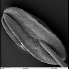
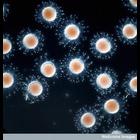
CIL:41308
NCBI Organism Classification
Penta lanceolata
Biological Process
anther development
Cellular Component
cell surface
Scanning electron microscope image of Penta lanceolata anther (pollen bearing part of the stamen).
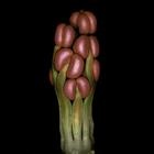
CIL:41814
NCBI Organism Classification
Spartium
Biological Process
flower formation
Cellular Component
cell surface
Spartium flower primordium (bud), captured using epi-illumination. Honorable Mention, 2010 Olympus BioScapes Digital Imaging Competition®.
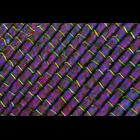
CIL:41832
NCBI Organism Classification
Elodea canadensis
Biological Process
none specified
Cellular Component
cell surface
Fresh water plant Elodea canadensis imaged using polarized light. 2010 Olympus BioScapes Digital Imaging Competition®.
CIL:42151
NCBI Organism Classification
Mus musculus
Biological Process
dissemination
Cellular Component
cell surface
Representative time-lapse movie of a normal mouse mammary fragment in collagen I. CIL 42168 is a related movie of a normal mammary fragment in Matrigel. Images taken every 20 min. This movie is par...
CIL:41558
NCBI Organism Classification
hydroid
Biological Process
polyp stage of life-cycle
Cellular Component
cell surface
Hydroid collected from kelp sample captured using epi-illumination and an image stack. Honorable Mention, 2011 Olympus BioScapes Digital Imaging Competition®.
CIL:25861
NCBI Organism Classification
Mus musculus
Biological Process
cell adhesion
Cellular Component
external side of plasma membrane
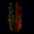
Movie shows restricted distribution of PECAM-1[high] CD45+ cells to the luminal surface of the aortic endothelium from an E11.5 mouse embryo. PECAM-1[high] cells are in red, CD45+ cells are in green a...
CIL:37314
NCBI Organism Classification
Cavia porcellus
Biological Process
none specified
Cellular Component
cell surface
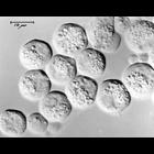
Nomaski image of live guinea pig pancreatic acinar cells. Image corresponds to Figure 1 in Proc Natl Acad Sci U S A. 1972 Oct;69(10):3028-32. Image made available by James D. Jamieson and the Departm...
CIL:37315
NCBI Organism Classification
Cavia porcellus
Biological Process
none specified
Cellular Component
cell surface
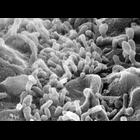
Scanning electron micrograph of single guinea pig pancreatic acinar cell. Image made available by James D. Jamieson and the Department of Cell Biology, Yale University School of Medicine.
CIL:39088
NCBI Organism Classification
Mus musculus
Biological Process
liver trabeculae organization
Cellular Component
cell surface
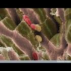
This scanning electron micrograph shows the internal structure of liver tissue from an adult mouse. The sinusoids (vascular channels lined with endothelial cells) can be seen as pink structures runnin...
CIL:39466
NCBI Organism Classification
Ascidiacea
Biological Process
embryo development
Cellular Component
cell surface
Early ascidian (sea squirt) embryos visualized by differential interface contrast (DIC) microscopy. Ascidians are used as a model for developmental research. Their simple embryonic development is rapi...
« Previous
1
...
3
4
5
6
7
8
9
10
...
138
Next »
Results per page:
10
20
50
100