Alternate header for print version
Contributors
Help
Submit
Search
menu
Data sets
Videos
Latest data
Center for Research in Biological Systems
Basic Science Building, Room 1000
University of California, San Diego
9500 Gilman Drive
La Jolla, CA 92093-0608, USA
Voice
: (858) 534-0276
Fax
: (858) 534-7497
Email
: dorloff@ncmir.ucsd.edu
Search Results for
cell periphery
(975 results)
CIL:40901
NCBI Organism Classification
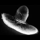
Didinium nasutum
Biological Process
phagocytosis
Cellular Component
oral apparatus
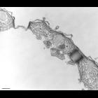
Didinium captures Paramecium. The cytostome opens at the end of the proboscis to accept its prey. Also showing metachronous waves of cilia within the two characteristic ciliary girdles of Didinium nas...
CIL:40951
NCBI Organism Classification
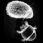
Didinium nasutum
Biological Process
phagocytosis
Cellular Component
oral apparatus
Didinium captures Paramecium. A moment after initial contact when toxicysts enter Paramecium. The strand of toxicysts can be seen between the two organisms, and Didinium will use the anchored toxicyst...
CIL:7598
NCBI Organism Classification
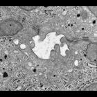
Cavia porcellus
Biological Process
canalicular bile acid transport
Cellular Component
tight junction
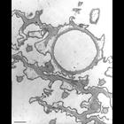
This is a view of apical domains of two adjacent hepatocytes showing the junctional complexes that attach cells to one another. The canaliculus is defined by the cell membranes of two adjacent liver c...
CIL:7599
NCBI Organism Classification
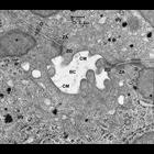
Cavia porcellus
Biological Process
canalicular bile acid transport
Cellular Component
tight junction
This is a view of apical domains of two adjacent hepatocytes in the guinea pig liver, showing the junctional complexes that attach cells to one another. The bile canaliculus (BC) is defined by the cel...
CIL:12604
NCBI Organism Classification
Paramecium multimicronucleatum
Biological Process
microtubule cytoskeleton organization
Cellular Component
cytoproct
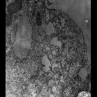
High resolution image of a colchicine-treated cell, used in an attempt to disrupt its microtubules. We found that microtubules already formed are not disrupted in Paramecium multimicronucleatum. This ...
CIL:9016
NCBI Organism Classification
Coleps hirtus
Biological Process
ciliary or flagellar motility
Cellular Component
cortical cytoskeleton
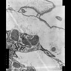
A high resolution image of a cortical monokinetid showing the basal body as well as postciliary and transverse microtubular ribbons. Slender endosomal vesicles accumulate near the bottom of a parasoma...
CIL:9701
NCBI Organism Classification
Coleps hirtus
Biological Process
water homeostasis
Cellular Component
contractile vacuole pore
Oblique section of the Coleps contractile vacuole pore revealing the supporting microtubules and adjacent fibrous layer. Standard glutaraldehyde fixation followed by osmium tetroxide, dehydrated in al...
CIL:12350
NCBI Organism Classification
Paramecium multimicronucleatum
Biological Process
defecation
Cellular Component
cytoproct
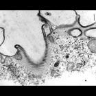
High resolution image of the somatic basal bodies along the sides of the cytoproct ridge have bundles of microtubules extending into the endoplasm from near their proximal ends. These bundles make con...
CIL:36233
NCBI Organism Classification
Tetrahymena pyriformis
Biological Process
contractile vacuole organization
Cellular Component
contractile vacuole pore
The two CV pores of a cell. An impression of the helically wound microtubules are detectable in the right pore. TEM taken on 4/21/78 by R. Allen with Hitachi HU11A operating at 60kV. Neg. 21,750X. Bar...
CIL:36758
NCBI Organism Classification
Paramecium caudatum
Biological Process
contractile vacuole organization
Cellular Component
contractile vacuole pore
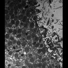
A circular depression on the dorsal surface of the cell marks the expulsion site of a contractile vacuole. This is referred to as the contractile vacuole “pore.". Microtubules encircle the pore. Tr...
« Previous
1
...
6
7
8
9
10
11
12
13
...
98
Next »
Results per page:
10
20
50
100