Alternate header for print version
Contributors
Help
Submit
Search
menu
Data sets
Videos
Latest data
Center for Research in Biological Systems
Basic Science Building, Room 1000
University of California, San Diego
9500 Gilman Drive
La Jolla, CA 92093-0608, USA
Voice
: (858) 534-0276
Fax
: (858) 534-7497
Email
: dorloff@ncmir.ucsd.edu
Search Results for
cell periphery
(975 results)
CIL:34732
NCBI Organism Classification
Tetrahymena pyriformis
Biological Process
cortical cytoskeleton organization
Cellular Component
cell cortex
Mucocysts have a herringbone pattern of subunits and are surrounded by a membrane that fuses with the plasma membrane. When the mucocyst content is extruded the content expands (see Hausmann, Protisto...
CIL:12111
NCBI Organism Classification
Colpoda cucullus
Biological Process
cortical cytoskeleton organization
Cellular Component
cell cortex
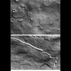
A micrograph showing the partially open cytoproct of Colpoda cucullus. The alveoli stop at the edges of the cytoproct and filamentous material (actin?) coats the inside of the single membrane that cov...
CIL:25541
NCBI Organism Classification
Drosophila melanogaster
Biological Process
calcium-dependent cell-cell adhesion
Cellular Component
adherens junction
A time-lapse movie showing the dorsal pouch formation and migration of heart-anchoring cells (HANCs) in the Drosophila embryo expressing ubi-DE-cad-GFP (Shg-GFP). Shg-positive HANCs corresponding to t...
CIL:10931
NCBI Organism Classification
Myotis
Biological Process
extracellular structure organization
Cellular Component
plasma membrane
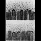
Filaments extend from microvilli of the epithelial cells in the intestine of the bat, Moytis lucifugus in these two high magnification electron micrographs. These filaments, which are 2.5-5nm thick, ...
CIL:11237
NCBI Organism Classification
Macaca mulatta
Biological Process
cell communication
Cellular Component
gap junction
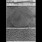
Representative examples of gap junctions from vertebrates (ciliary epithelium from the eye of a Macaca mulatta, upper) and invertebrates (inverted gap junctions from cells of the mid-gut of the horses...
CIL:11221
NCBI Organism Classification
Carassius auratus
Biological Process
cell communication
Cellular Component
gap junction
Typical organization of gap junctions as seen in thin sections (upper) and replicas of freeze-fractured cell membranes (middle and lower). Upper, gap junction from the saccular macula of a goldfish; ...
CIL:22782
NCBI Organism Classification
Didinium nasutum
Biological Process
phagocytosis
Cellular Component
oral apparatus
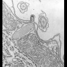
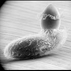
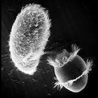
Didinium intiates capture of Paramecium. Believe-it-or-not, this Didinium may accomplish the ingestion of the Paramecium in less than one minute. See other images in the link below. Also showing metac...
CIL:21992
NCBI Organism Classification
Didinium nasutum
Biological Process
phagocytosis
Cellular Component
oral apparatus
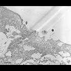
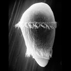
Didinium ingests Paramecium. Note that Paramecium is nearly the waiting food vacuole and the proboscis of Didinium is beginning to reform. This micrograph also shows the metachronous waves of cilia in...
CIL:21995
NCBI Organism Classification
Didinium nasutum
Biological Process
phagocytosis
Cellular Component
oral apparatus
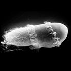
Didinium ingests Paramecium. Note that Paramecium is folded in half as it is compressed and enters the waiting food vacuole. This micrograph also shows a few discharged Paramecium trichocysts as well ...
CIL:21993
NCBI Organism Classification
Didinium nasutum
Biological Process
phagocytosis
Cellular Component
oral apparatus
Didinium captures Paramecium. A moment after initial contact when toxicysts enter Paramecium. The strand of toxicysts can be seen between the two organisms, and Didinium will use the anchored toxicyst...
« Previous
1
...
5
6
7
8
9
10
11
12
...
98
Next »
Results per page:
10
20
50
100