Alternate header for print version
Contributors
Help
Submit
Search
menu
Data sets
Videos
Latest data
Center for Research in Biological Systems
Basic Science Building, Room 1000
University of California, San Diego
9500 Gilman Drive
La Jolla, CA 92093-0608, USA
Voice
: (858) 534-0276
Fax
: (858) 534-7497
Email
: dorloff@ncmir.ucsd.edu
Search Results for
cell periphery
(975 results)
CIL:17447
NCBI Organism Classification
Paramecium multimicronucleatum
Biological Process
clathrin coating of Golgi vesicle, plasma membrane to endosome targeting
Cellular Component
cell cortex
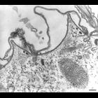
Cell sectioned perpendicular to the surface. The parasomal sac ends internally as a coated pit, coated with clathrin. Where the coated pit has pinched off a clathrin-coated vesicle is formed. When the...
CIL:11225
NCBI Organism Classification
none specified
Biological Process
cell communication
Cellular Component
gap junction
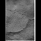
Replica of a freeze-fractured gap junction presents the inner half-membrane on one of the cells (P-face, lower half of the micrograph) and the the outer half-membrane of the other cells (E-face, upper...
CIL:11230
NCBI Organism Classification
Rattus
Biological Process
cell communication
Cellular Component
gap junction
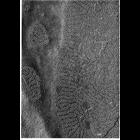
Gap junctions from a granulosa cell of a rat ovarian follicle. Variability in size of the gap junctions is likely an artifact of the freeze fracture preparation. Figure 99 from Chapter 3 (Junctional ...
CIL:11232
NCBI Organism Classification
none specified
Biological Process
cell communication
Cellular Component
gap junction
Gap junctions in ciliary epithelium under conditions of oxygenation (upper) and anoxia (lower), in tissue prepared by ultrarapid freezing using liquid helium. Under oxygenated conditions, the connexo...
CIL:11923
NCBI Organism Classification
Rattus
Biological Process
none specified
Cellular Component
clathrin coat of trans-Golgi network vesicle
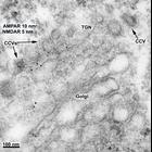
NMDA receptors (NMDARs) can exit the Golgi/TGN via clathrin-coated vesicles. Double-labeling using rabbit polyclonal antibodies to AMPA receptors (AMPARs) (combination of 3 antibodies: GluR1, GluR2, G...
CIL:10929
NCBI Organism Classification
Felis catus
Biological Process
extracellular structure organization
Cellular Component
plasma membrane
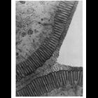
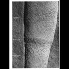
This electron micrograph shows a region of the brush border of the cat intestine. Tufted and branched polysaccharide filaments, each a few nanometers thick, extend from the microvilli to make up the ...
CIL:10932
NCBI Organism Classification
Felis catus
Biological Process
extracellular structure organization
Cellular Component
plasma membrane
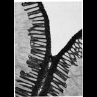
This electron micrograph highlights a darkly-stained glycocalyx rim of the brush border of the intestinal epithelium of the cat, stained en bloc with colloidal thorium. The glycocalyx is composed of ...
CIL:17892
NCBI Organism Classification
Didinium nasutum
Biological Process
phagocytosis
Cellular Component
oral apparatus
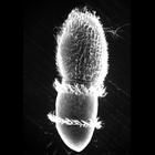
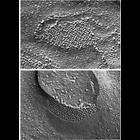
Didinium ingests Paramecium. Also showing a few discharged trichocysts and metachronous waves of cilia in the two characteristic ciliary girdles of Didinium nasutum. This micrograph was taken in 1968 ...
CIL:10900
NCBI Organism Classification
unidentified monkey
Biological Process
plasma membrane organization
Cellular Component
plasma membrane
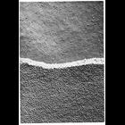
This electron micrograph shows the E-fracture face (above) and P-fracture face (below) of the membrane of adjacent epithelial cells from the ciliary epithelium of the monkey eye. Intramembranous part...
CIL:10903
NCBI Organism Classification
Cavia porcellus
Biological Process
fertilization
Cellular Component
plasma membrane
This image, produced by the freeze-fracture technique, shows regional specialization in the structure of mammalian sperm. The P-face of the plasma membrane of a guinea pig sperm tail can be divided i...
« Previous
1
...
3
4
5
6
7
8
9
10
...
98
Next »
Results per page:
10
20
50
100

")