Alternate header for print version
Contributors
Help
Submit
Search
menu
Data sets
Videos
Latest data
Center for Research in Biological Systems
Basic Science Building, Room 1000
University of California, San Diego
9500 Gilman Drive
La Jolla, CA 92093-0608, USA
Voice
: (858) 534-0276
Fax
: (858) 534-7497
Email
: dorloff@ncmir.ucsd.edu
Search Results for
adherens junction
(48 results)
CIL:10105
NCBI Organism Classification
Homo sapiens
Biological Process
none specified
Cellular Component
clathrin coat of coated pit
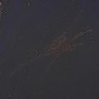
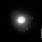
Human retinal pigmented epithelial (RPE) cells labeled for clathrin-coated pits (green), focal adhesions (red) and nuclei (blue). RPE cells stabily expressing 'clathrin light chain a' tagged with EGFP...
CIL:12598
NCBI Organism Classification
Canis lupus familiaris
Biological Process
none specified
Cellular Component
stress fiber
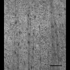
Parallel stress fibers on the ventral face of an MDCK cell, just inside the plasma membrane. This transmission electron micrograph of a flat-embedded MDCK cell was taken 60-70nm from the interface bet...
CIL:25702
NCBI Organism Classification
Rattus
Biological Process
tyrosine phosporylation
Cellular Component
focal adhesion
MTLn3 cell cotransfected with FAK siRNA (target A) and two tandem Src SH2 phosphotyrosine-binding domains (YFP-dSH2), which specifically detects tyrosine phosphorylation at focal adhesions. CIL:25703...
CIL:38652
NCBI Organism Classification
Homo sapiens
Biological Process
molecular organization
Cellular Component
focal adhesion
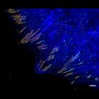
This image combines total internal reflection microscopy (TIRF) of mCerulean-actin (blue) with photoactivation localization microscopy (PALM) image of tdEos-vinculin (red) and Dronpa-paxillin (green)....
CIL:38600
NCBI Organism Classification
Homo sapiens
Biological Process
molecular organization
Cellular Component
focal adhesion
Photoactivation localization microscopy image (PALM) of a human foreskin fibroblast expressing Dronpa-actin (green) and tdEos-paxillin (red). Paxillin assembles in fibrillar-like adhesions that run p...
CIL:40460
NCBI Organism Classification
Homo sapiens
Biological Process
organelle organization
Cellular Component
focal adhesion
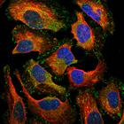
Single slice confocal images representing one optical slice of a cell stained for the Human Protein Atlas (HPA) antibody HPA005724 (shown in green). HPA005724 recognizes VASP (vasodilator-stimulated ...
CIL:7440
NCBI Organism Classification
Homo sapiens
Biological Process
regulation of focal adhesion assembly
Cellular Component
focal adhesion
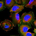
Human umbilical vein endothelial cells (HUVECS) labeled for focal adhesions (green) and F-actin (red). HUVECs were plated on polyacrilamide gels (1.5 Kpa) coated with 1.0 mg/ml fibronectin. After 2...
CIL:12285
NCBI Organism Classification
Mus musculus
Biological Process
migration in 3D matrix
Cellular Component
focal adhesion
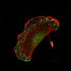
This confocal image of beta3-/- integrin cells plated on cell-derived matrices has an accompanying image of the wild type control cells. There are ncreased numbers of peripheral matrix-associated adh...
CIL:12286
NCBI Organism Classification
Mus musculus
Biological Process
migration in 3D matrix
Cellular Component
focal adhesion
This confocal image of wild type cells plated on cell-derived matrices that correspond to an accompanying image of beta3-/- integrin cells. There are ncreased numbers of peripheral matrix-associated ...
CIL:41674
NCBI Organism Classification
Homo sapiens
Biological Process
organelle organization
Cellular Component
focal adhesion
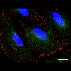
Single slice confocal images representing one optical slice of a cell stained for the Human Protein Atlas (HPA) antibody HPA001842 (shown in green). HPA001842 is a polyclonal anti-PTK2 antibody that l...
« Previous
1
2
3
4
5
Next »
Results per page:
10
20
50
100


")