Alternate header for print version
Contributors
Help
Submit
Search
menu
Data sets
Videos
Latest data
Center for Research in Biological Systems
Basic Science Building, Room 1000
University of California, San Diego
9500 Gilman Drive
La Jolla, CA 92093-0608, USA
Voice
: (858) 534-0276
Fax
: (858) 534-7497
Email
: dorloff@ncmir.ucsd.edu
Search Results for
adherens junction
(48 results)
CIL:38651
NCBI Organism Classification
Homo sapiens
Biological Process
molecular organization
Cellular Component
focal adhesion
This photoactivation localization microscopy (PALM) image of tdEos-vinculin (red) and Dronpa-paxillin (green) illustrates that vinculin and paxillin are segregated into interlocking microdomains withi...
CIL:132
NCBI Organism Classification
Mus musculus
Biological Process
regulation of actin cytoskeleton organization
Cellular Component
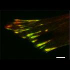
cell leading edge
NIH 3T3 cell transfected with EGFP-VASP. VASP is localized to the focal adhesions and is also present along the protruding leading edge. VASP only hightlights the portions of the periphery that are ...
CIL:38599
NCBI Organism Classification
Homo sapiens
Biological Process
focal adhesion assembly
Cellular Component
focal adhesion
This total internal reflection (TIRF) image of both the Dronpa-alphaa ctinin and the unconverted tdEos-vinculin channels corresponds to the same image field as the diffraction limited DIC image CIL 38...
CIL:25846
NCBI Organism Classification
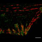
Cricetulus griseus
Biological Process
focal adhesion disassembly
Cellular Component
focal adhesion
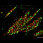
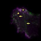
Time-lapse of a CHO.K1 cell cotransfected with very low levels of GFP-MIIA (myosin IIA, green) and paxillin (magenta). Yellow arrowheads mark paxillin-containing adhesions that disassemble concomitant...
CIL:11180
NCBI Organism Classification
Rattus
Biological Process
maintenance of apical/basal cell polarity
Cellular Component
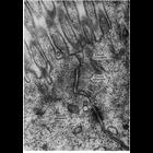
apical junction complex
Electron micrograph of the junctional complex of intestinal epithelial cells of the rat shows the apical-most zonula occludens (tight junction), the zonula adherens (medium junction) and the macula ad...
CIL:38602
NCBI Organism Classification
Homo sapiens
Biological Process
molecular organization
Cellular Component
focal adhesion
This photoactivation localization microscopy (PALM) image of tdEos-paxillin (green) and PsCFP20-zyxin (red) demonstrates that these two focal adhesion proteins are have very little over-lap when visua...
CIL:25703
NCBI Organism Classification
Rattus
Biological Process
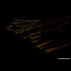
tyrosine phosphorylation
Cellular Component
focal adhesion
MTLn3 cell cotransfected with control siRNA and two tandem Src SH2 phosphotyrosine-binding domains (YFP-dSH2), which specifically detects tyrosine phosphorylation at focal adhesions. CIL:25702 is cor...
CIL:38598
NCBI Organism Classification
Homo sapiens
Biological Process
molecular organization of focal adhesion components
Cellular Component
focal adhesion
Photoactivation localization microscopy image (PALM) of a human foreskin fibroblast expressing Dronpa-alpha-actinin (red) and tdEos-vinculin (green). This image reveals that although conventional dif...
CIL:38604
NCBI Organism Classification
Homo sapiens
Biological Process
molecular organization
Cellular Component
focal adhesion
This total internal reflection (TIRF) image of unconverted tdEos-paxillin (green) and PsCFP20-zyxin (red) demonstrates that these two proteins are almost completely colocalized when observed with diff...
CIL:7439
NCBI Organism Classification
Mus musculus
Biological Process
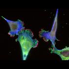
cell adhesion
Cellular Component
focal adhesion
Mouse Embryonic Fibroblasts (MEFs) grown on glass coverslips coated with 10 ug/ml Fibronectin. After 24 hrs cells were fixed in 3% paraformaldehyde and stained with mouse antibody to Paxillin, rabbit...
« Previous
1
2
3
4
5
Next »
Results per page:
10
20
50
100