Alternate header for print version
Contributors
Help
Submit
Search
menu
Data sets
Videos
Latest data
Center for Research in Biological Systems
Basic Science Building, Room 1000
University of California, San Diego
9500 Gilman Drive
La Jolla, CA 92093-0608, USA
Voice
: (858) 534-0276
Fax
: (858) 534-7497
Email
: dorloff@ncmir.ucsd.edu
Search Results for
skeletal muscle cell
(140 results)
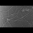
CIL:6263
NCBI Organism Classification
Oryctolagus cuniculus
Biological Process
skeletal muscle contraction
Cellular Component
cytoskeleton
Rabbit psoas skeletal muscle fibers were blended in the presence of 5 mM MgATP to dissociate thick and thin filaments. The medium was then gently replaced with 0.3M KCl to dissociate thick filaments....
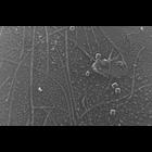
CIL:6267
NCBI Organism Classification
Oryctolagus cuniculus
Biological Process
skeletal muscle contraction
Cellular Component
cytoskeleton
Rabbit psoas skeletal muscle fibers were blended in the presence of 5 mM MgATP to dissociate thick and thin filaments. The medium was then gently replaced with 0.3M KCl to dissociate thick filaments....
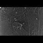
CIL:831
NCBI Organism Classification
Oryctolagus cuniculus
Biological Process
skeletal muscle contraction
Cellular Component
cytoskeleton
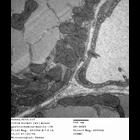
Rabbit skeletal muscle fibers were minced in a ATP-containing relaxing buffer and dissociated into thick and thin filaments by mild blending. The isolated filaments were then deposited onto mica flak...
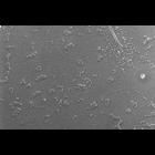
CIL:1435
NCBI Organism Classification
Oryctolagus cuniculus
Biological Process
skeletal muscle contraction
Cellular Component
cytoskeleton
Rabbit psoas skeletal muscle fibers were blended in the presence of 5 mM MgATP to dissociate thick and thin filaments. The medium was then gently replaced with 0.3M KCl to dissociate thick filaments....
CIL:258
NCBI Organism Classification
Mus musculus
Biological Process
plasma membrane organization
Cellular Component
dystroglycan complex
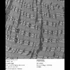
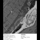
Thin section electron microscopy of diaphragm (skeletal) muscle from a wild type mouse. Sample was viewed with a Hitachi 7600 electron microscope (accelerating voltage 80 KV) and imaged with an AMT d...
CIL:266
NCBI Organism Classification
Mus musculus
Biological Process
plasma membrane organization
Cellular Component
dystroglycan complex
Thin section electron microscopy of diaphragm (skeletal) muscle from a wild type mouse. Sample was viewed with a Hitachi 7600 electron microscope (accelerating voltage 80 KV) and imaged with an AMT d...
CIL:339
NCBI Organism Classification
Mus musculus
Biological Process
plasma membrane organization
Cellular Component
dystroglycan complex
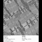
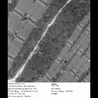
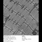
Thin section electron microscopy of gastrocnemius (skeletal) muscle from a wild type mouse. Sample was viewed with a Hitachi 7600 electron microscope (accelerating voltage 80 KV) and imaged with an A...
CIL:345
NCBI Organism Classification
Mus musculus
Biological Process
plasma membrane organization
Cellular Component
dystroglycan complex
Thin section electron microscopy of gastrocnemius (skeletal) muscle from a wild type mouse. Sample was viewed with a Hitachi 7600 electron microscope (accelerating voltage 80 KV) and imaged with an A...
CIL:346
NCBI Organism Classification
Mus musculus
Biological Process
plasma membrane organization
Cellular Component
dystroglycan complex
Thin section electron microscopy of gastrocnemius (skeletal) muscle from a wild type mouse. Sample was viewed with a Hitachi 7600 electron microscope (accelerating voltage 80 KV) and imaged with an A...
CIL:342
NCBI Organism Classification
Mus musculus
Biological Process
plasma membrane organization
Cellular Component
dystroglycan complex
Thin section electron microscopy of gastrocnemius (skeletal) muscle from a wild type mouse. Sample was viewed with a Hitachi 7600 electron microscope (accelerating voltage 80 KV) and imaged with an A...
« Previous
1
...
6
7
8
9
10
11
12
13
14
Next »
Results per page:
10
20
50
100