Alternate header for print version
Contributors
Help
Submit
Search
menu
Data sets
Videos
Latest data
Center for Research in Biological Systems
Basic Science Building, Room 1000
University of California, San Diego
9500 Gilman Drive
La Jolla, CA 92093-0608, USA
Voice
: (858) 534-0276
Fax
: (858) 534-7497
Email
: dorloff@ncmir.ucsd.edu
Search Results for
myoblast
(1735 results)
CIL:39086
NCBI Organism Classification
none specified
Biological Process
none specified
Cellular Component
myofibril
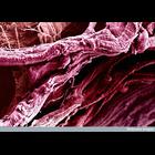
Colorized scanning electron micrograph of thigh muscle fibrils.
CIL:11430
NCBI Organism Classification
Felis catus
Biological Process
cellular respiration
Cellular Component
mitochondrion
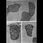
Figure 239 from Chapter 7 (Mitochondria) of 'The Cell, 2nd Ed.' by Don W. Fawcett M.D. The cristae of mitochondria can take on zig-zag configuration (see arrows) which is particularly apparent in cel...
CIL:37202
NCBI Organism Classification
Cavia porcellus
Biological Process
none specified
Cellular Component
nucleus
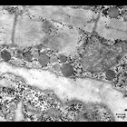
Transmission electron micrograph of a section of the right atrium of a guinea pig heart. A portion of nucleus is seen at far left, with two centrioles close to the nuclear envelope. A region of myofib...
CIL:37207
NCBI Organism Classification
Rattus
Biological Process
regulation of cardiac muscle contraction
Cellular Component
intercalated disc
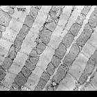
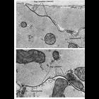
Transmission electron micrograph of longitudinal section of rat heart cardiac muscle from the atrium. A darkly stained intercalated disc runs from upper left to lower right. Also prominent are groups...
CIL:37209
NCBI Organism Classification
Rattus
Biological Process
regulation of cardiac muscle contraction
Cellular Component
atrial granuales
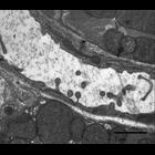
Transmission electron micrograph of longitudinal section of rat cardiac muscle from the heart atrium. Homogeneously staining spherical trial granules are present as well as mitochondria and myofibrils...
CIL:37212
NCBI Organism Classification
Rattus
Biological Process
regulation of cardiac muscle contraction
Cellular Component
mitochondrion
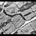
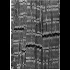
Transmission electron micrograph of longitudinal section of a a rat heart ventricle. Ordered arrays of myofibrils are interspersed with rows of mitochondria. Small, darkly staining glycogen granules a...
CIL:39754
NCBI Organism Classification
Mus musculus
Biological Process
transport
Cellular Component
none specified
Transmission electron micrograph of mouse cardiac muscle. Transport is visible in the ventricle across a space from one muscle cell to the adjacent muscle cell.
CIL:11243
NCBI Organism Classification
Felis catus
Biological Process
cell communication
Cellular Component
gap junction
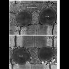
Papillary muscle from cat heart shows a step-like end-to-end junction of two cardiac muscle cells. The longitudinally oriented lateral cell boundaries are straight and exhibit extensive gap junctions...
CIL:11245
NCBI Organism Classification
Felis catus
Biological Process
cell communication
Cellular Component
gap junction
Gap junctions in papillary muscle from cat heart. These communicating junctions are responsible for the spreading wave of depolarization through the myocardium. Desmosomes can also be found on the l...
CIL:11456
NCBI Organism Classification
Felis catus
Biological Process
cellular respiration
Cellular Component
mitochondrion
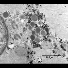
Lipid droplets partially enveloped by mitochondria in the interstices between cardiac myofibrils reveal a metabolic partnership that can serve as an alternative energy source for muscle. Figures 255 ...
« Previous
1
2
3
4
5
6
7
8
9
...
174
Next »
Results per page:
10
20
50
100