Alternate header for print version
Contributors
Help
Submit
Search
menu
Data sets
Videos
Latest data
Center for Research in Biological Systems
Basic Science Building, Room 1000
University of California, San Diego
9500 Gilman Drive
La Jolla, CA 92093-0608, USA
Voice
: (858) 534-0276
Fax
: (858) 534-7497
Email
: dorloff@ncmir.ucsd.edu
Search Results for
fibroblast
(195 results)
CIL:36073
NCBI Organism Classification
Mus musculus
Biological Process
actin cytoskeleton organization
Cellular Component
actin cytoskeleton
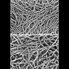
Figures 442 (upper) and 443 (lower) from Chapter 16 (Cytoplasmic matrix and cytoskeleton) of 'The Cell, 2nd Ed.' by Don W. Fawcett M.D. Purified preparation of actin undecorated (upper) and decorated...
CIL:36031
NCBI Organism Classification
Homo sapiens
Biological Process
none specified
Cellular Component
nuclear lamina
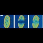
To measure the in vivo mobility of the major nuclear lamina protein lamin A, human fibroblasts were transfected with the gene fused to EGFP. Fluorescence images were recorded with a confocal microsco...
CIL:38985
NCBI Organism Classification
Rattus
Biological Process
cell adhesion
Cellular Component
stress fiber
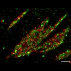
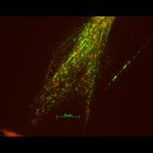
Fluorescent micrograph of rat embryo fibroblast cell growing in serum stained to reveal actin stress fibres (red) and vinculin (component of focal adhesions) in green/yellow.
CIL:38651
NCBI Organism Classification
Homo sapiens
Biological Process
molecular organization
Cellular Component
focal adhesion
This photoactivation localization microscopy (PALM) image of tdEos-vinculin (red) and Dronpa-paxillin (green) illustrates that vinculin and paxillin are segregated into interlocking microdomains withi...
CIL:40687
NCBI Organism Classification
Mus musculus
Biological Process
chromosome organization
Cellular Component
nuclear chromosome
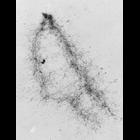
High voltage (1MeV) transmission electron microscopy image of an isolated metaphase chromatid pair from a mouse A9 fibroblast, showing the fiber-like structure.
CIL:9070
NCBI Organism Classification
Rattus
Biological Process
mitochondrion organization
Cellular Component
mitochondrion
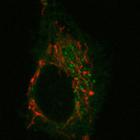
NRK cells expressing a mitochondria marker, mito-RFP (red), and PSS1-CFP (green), phosphatidylserine synthase 1. A single 6 micron confocal slice was imaged on a Zeiss LSM 510 every 5.9 sec with the...
CIL:10276
NCBI Organism Classification
Mus musculus
Biological Process
platelet-derived growth factor receptor signaling pathway
Cellular Component
dynamin
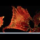
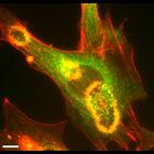
After over-night serum starvation, mouse fibroblast expressing Dyn2-GFP were stimulated with 10ng/ml PDGF. After 5min, the cells were fixed and stained with rhodamine-phalloidin. PDGF-induced dorsal m...
CIL:12625
NCBI Organism Classification
Homo sapiens
Biological Process
stem cell proliferation
Cellular Component
cell
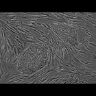
Human embryonic stem cell colonies. This phase contrast image shows two human embryonic stem cell colonies growing on a feeder layer of mouse embryonic fibroblasts. The stem cells actually grow in bet...
CIL:245
NCBI Organism Classification
Mus musculus
Biological Process
intermediate filament-based process
Cellular Component
mitochondrion
Mitochondrial distribution (visualized with Mitotracker CMXRos) was largely unaffected by partial fragmentation of the intermediate filament cytoskeleton, visualized here with vimentin antibody, after...
CIL:23041
NCBI Organism Classification
Rattus
Biological Process
gelsolin treatment
Cellular Component
intermediate filament
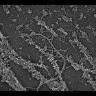
Association of plectin with myosin II. A gelsolin-treated REF-52 cytoskeleton immunogold labeled myosin II (10 nm). After actin depletion, intermediate filaments with plectin sidearms remain associate...
« Previous
1
2
3
4
5
6
7
8
9
...
20
Next »
Results per page:
10
20
50
100