Alternate header for print version
Contributors
Help
Submit
Search
menu
Data sets
Videos
Latest data
Center for Research in Biological Systems
Basic Science Building, Room 1000
University of California, San Diego
9500 Gilman Drive
La Jolla, CA 92093-0608, USA
Voice
: (858) 534-0276
Fax
: (858) 534-7497
Email
: dorloff@ncmir.ucsd.edu
Search Results for
endocrine cell
(38 results)
CIL:35981
NCBI Organism Classification
Spermophilus citellus
Biological Process
lipid storage
Cellular Component
lipid particle
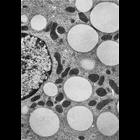
Figure 360 from Chapter 15 (Cytoplasmic Inclusions) of 'The Cell, 2nd Ed.' by Don W. Fawcett M.D. Leydig cell from a reproductively active ground squirrel, Citellus lateralis. Physiological state ca...
CIL:11105
NCBI Organism Classification
Mus musculus
Biological Process
plasma membrane organization
Cellular Component
cell surface
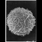
Scanning electron micrograph of the surface of a peritoneal mast cell from mouse shows undulating folds, and few microvilli. The membrane folds could be misinterpreted for microvilli if analyzed in t...
CIL:11439
NCBI Organism Classification
Spermophilus citellus
Biological Process
cellular respiration
Cellular Component
mitochondrion
Mitochondria can show tremendous variation in size, even within a single cell. Here, a huge spherical mitochondria of about 6-7 µm in diameter is adjacent to small elongate mitochondria 0.5 µm wide...
CIL:41111
NCBI Organism Classification
Cavia porcellus
Biological Process
organelle organization
Cellular Component
nucleus
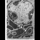
A transmission electron micrograph of a Leydig cell isolated from guinea pig testes.The eccentric nucleus and several pale lipid droplets are visible. Numerous mitochondria and smooth endoplasmic ret...
CIL:11384
NCBI Organism Classification
Cavia porcellus
Biological Process
post-translational protein modification
Cellular Component
Golgi apparatus
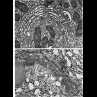
Figures 213 (upper) and 214 (lower) from Chapter 6 (Golgi Apparatus) of 'The Cell, 2nd Ed.' by Don W. Fawcett M.D. The Golgi complex of a Leydig cell from guinea pig testis (upper) and from and acina...
CIL:41355
NCBI Organism Classification
Cavia porcellus
Biological Process
organelle organization
Cellular Component
nucleolus
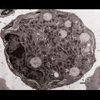
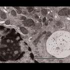
A transmission electron micrograph of a Leydig cell isolated from guinea pig testes. This is a higher magnification image of CIL: 41116 highlighting the distinct nucleolus and a membrane-bound, debri...
CIL:11048
NCBI Organism Classification
Cavia porcellus
Biological Process
nucleus organization
Cellular Component
nuclear envelope
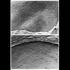
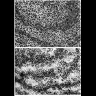
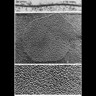
Nuclear pores are clearly seen in these transmission electron micrographs of cells after cryo-fixation, freeze-fracture and surface replication. Upper panel shows the outer surface of of the nuclear e...
CIL:11437
NCBI Organism Classification
Phodopus
Biological Process
cellular respiration
Cellular Component
mitochondrion
Tubules within the cristae of mitochondria can sometimes take on unusual configurations. Here, in the upper panel, a mitochondrion from an astrocyte in hamster brain appears to have prismatic tubules...
CIL:10772
NCBI Organism Classification
Homo sapiens
Biological Process
translation
Cellular Component
endoplasmic reticulum
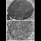
Figures 173 (upper panel) and 174 (lower panel) from Chapter 5 (Endoplasmic Reticulum) of 'The Cell, 2nd Ed.' by Don W. Fawcett M.D. Electron micrographs of endoplasmic reticulum with the section pla...
CIL:11221
NCBI Organism Classification
Carassius auratus
Biological Process
cell communication
Cellular Component
gap junction
Typical organization of gap junctions as seen in thin sections (upper) and replicas of freeze-fractured cell membranes (middle and lower). Upper, gap junction from the saccular macula of a goldfish; ...
« Previous
1
2
3
4
Next »
Results per page:
10
20
50
100