Alternate header for print version
Contributors
Help
Submit
Search
menu
Data sets
Videos
Latest data
Center for Research in Biological Systems
Basic Science Building, Room 1000
University of California, San Diego
9500 Gilman Drive
La Jolla, CA 92093-0608, USA
Voice
: (858) 534-0276
Fax
: (858) 534-7497
Email
: dorloff@ncmir.ucsd.edu
Search Results for
Outer plexiform layer of retina
(14811 results)
CIL:41630
NCBI Organism Classification
Mus musculus
Biological Process
none specified
Cellular Component
cell projection
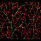
Confocal micrograph of a mouse retina showing retinal astrocytes in red and blood vessels in green. Honorable Mention, 2011 Olympus BioScapes Digital Imaging Competition®.
CCDB:54
Species
mouse
Organ
eye
Cell type
photoreceptor/cone
System
central nervous system
Structure
mitochondrion
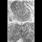
Cone and rod mitochondria: electron tomography
CIL:39788
NCBI Organism Classification
Lytechinus pictus
Biological Process
embryonic morphogenesis
Cellular Component
cell surface
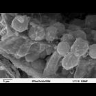
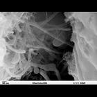
Scanning electron microscope image of Strongylocentrotus drobachiensus embryo at the primary mesenchyme blastula stage. Embryo was split open to reveal the outer epithelial layer and the blastocoel ca...
CIL:11418
NCBI Organism Classification
Talpidae
Biological Process
mitochondrial fission
Cellular Component
mitochondrion
Figure 232 from Chapter 7 (Mitochondria) of 'The Cell, 2nd Ed.' by Don W. Fawcett M.D. Dividing mitochondria in gasric mucosa of a mole. Upper panel, the bilaminar septum as formed, but the outer me...
CIL:10950
NCBI Organism Classification
Felis catus
Biological Process
plasma membrane organization
Cellular Component
basement membrane
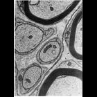
Schwann cells in peripheral nerves secrete a layer similar to the basal lamina called the lamina externa, boundary layer, or basement membrane. In this micrograph, the lamina externa surrounds the Sc...
CIL:39791
NCBI Organism Classification
Lytechinus pictus
Biological Process
embryonic morphogenesis
Cellular Component
extracellular matrix
Scanning electron microscope image of Strongylocentrotus drobachiensus [sea urchin] embryo at the late gastrula stage. This high magnification image of the embryo shows the primary mesenchyme syncytia...
CIL:35531
NCBI Organism Classification
Lilium longiflorum
Biological Process
pollination
Cellular Component
pollen wall
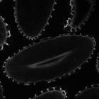
Lilium longiflorum (Easter Lily) pollen. This is a thin optical section through the center of the desiccated stage of the mature pollen showing autofluorescence or harmonic generation of intrinsic st...
CIL:40389
NCBI Organism Classification
Helianthus annuus
Biological Process
plant stem organization
Cellular Component
cortex
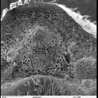
Scanning electron microscope image of cross-section through Helianthus annuus (sunflower) stem. The image shows the outer epidermal layer, followed by the cortex and then large vascular bundles. The v...
CIL:35529
NCBI Organism Classification
Lilium longiflorum
Biological Process
pollination
Cellular Component
pollen wall
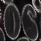
Lilium longiflorum (Easter Lily) pollen. This is a thin optical section through the center of the desiccated stage of the mature pollen showing autofluorescence or harmonic generation of intrinsic st...
CIL:35535
NCBI Organism Classification
Lilium longiflorum
Biological Process
pollination
Cellular Component
pollen wall
Lilium longiflorum (Easter Lily) pollen. This is a thin optical section through the center of the desiccated stage of the mature pollen showing autofluorescence or harmonic generation of intrinsic st...
« Previous
1
2
3
4
5
6
7
8
9
...
1482
Next »
Results per page:
10
20
50
100


")