Alternate header for print version
Contributors
Help
Submit
Search
menu
Data sets
Videos
Latest data
Center for Research in Biological Systems
Basic Science Building, Room 1000
University of California, San Diego
9500 Gilman Drive
La Jolla, CA 92093-0608, USA
Voice
: (858) 534-0276
Fax
: (858) 534-7497
Email
: dorloff@ncmir.ucsd.edu
Search Results for
Outer plexiform layer of retina
(14805 results)
CIL:11636
NCBI Organism Classification
Rattus sp.
Biological Process
epithelial cilium movement
Cellular Component
cilium
Figures 323 & 324 from Chapter 13 (Cilia and Flagella) of 'The Cell' by Don W. Fawcett M.D. The outer segments of the rods and cones of the vertebrate retina and many photoreceptors of invertebrates b...
CIL:38802
NCBI Organism Classification
Mus musculus
Biological Process
neural retina development
Cellular Component
nucleus
This confocal micrograph shows the detailed structure of the retina from a one-month-old mouse. The retina is the photoreceptive organ of the eye. It is composed of layers of neuronal cells that captu...
CIL:39745
NCBI Organism Classification
Mus musculus
Biological Process
photoreception
Cellular Component
nucleus
TEM image of epon section of a freeze-substituted mouse retina illustrating the unusual distribution of heterochromatin in rod receptor nuclei where the compact heterochromatin is centrally located. W...
CIL:39027
NCBI Organism Classification
Danio rerio
Biological Process
Dlx4/6 promotor specification
Cellular Component
neuron projection
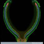
Confocal micrograph of the head region of a transgenic zebrafish embryo. The large circular structure is the eye, showing the structure of the retina. Some neurons in the brain are highlighted in gre...
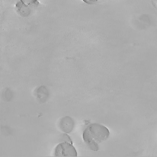
CCDB:7742
Species
mouse
Organ
eye
Cell type
none specified
System
central nervous system
Structure
none specified
Ultrastructural Characterization of the Mouse Optic nerve Head and Retina
CIL:39340
NCBI Organism Classification
Nicotiana alata
Biological Process
plant stem organization
Cellular Component
none specified
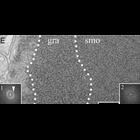
Scanning electron microscope image of Nicotiana alata stem cross section. Image shows outer epidermal layer, followed by the cortex and then large vascular bundles. The vascular bundles contain the ph...
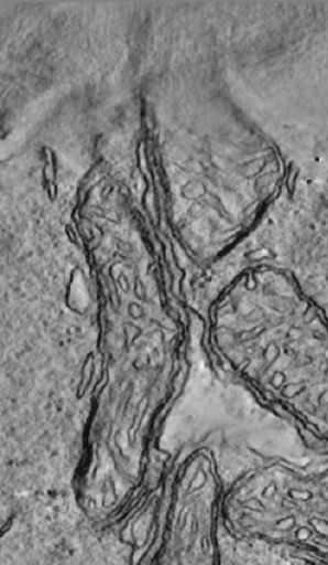
CCDB:8495
Species
Mouse
Organ
eye
Cell type
retina rod cell
System
central nervous system
Structure
rod spherule
Cone and rod mitochondria: electron tomography
CIL:40386
NCBI Organism Classification
Psidium guajava
Biological Process
plant stem organization
Cellular Component
mucilage
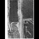
Scanning electron microscope image of cross-section through a Psidium guajava stem. The image shows the thin outer epidermal layer with a thick layer of cortex beneath. The vascular area consists of ...
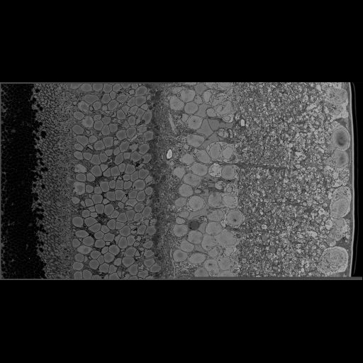
CCDB:8747
Species
mouse
Organ
eye
Cell type
retina rod cell
System
central nervous system
Structure
mitochondrion
Cone and rod mitochondria: electron tomography
CIL:38803
NCBI Organism Classification
Danio rerio
Biological Process
gene expression
Cellular Component
none specified
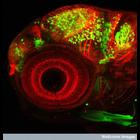
This photomicrograph shows the retina from the eye of a three-day-old zebrafish. The retina is viewed here from the front, as if the viewer is looking directly into the eye of the fish. This image is...
« Previous
1
2
3
4
5
6
7
8
9
...
1481
Next »
Results per page:
10
20
50
100