Alternate header for print version
Contributors
Help
Submit
Search
menu
Data sets
Videos
Latest data
Center for Research in Biological Systems
Basic Science Building, Room 1000
University of California, San Diego
9500 Gilman Drive
La Jolla, CA 92093-0608, USA
Voice
: (858) 534-0276
Fax
: (858) 534-7497
Email
: dorloff@ncmir.ucsd.edu
Search Results for
Outer nuclear layer of retina
(14816 results)
CIL:11812
NCBI Organism Classification
Danio rerio
Biological Process
anatomical structure morphogenesis
Cellular Component
nucleus
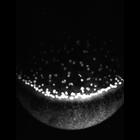
Timelapse shows yolk syncytial layer (YSL) nuclear migration on the ventral side during gastrulation. Divergence of YSL nuclei away from ventral midline can be seen. As epiboly ends and blastopore clo...
CIL:11813
NCBI Organism Classification
Danio rerio
Biological Process
anatomical structure morphogenesis
Cellular Component
nucleus
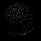
Timelapse of yolk syncytial layer (YSL) nuclear movements during gastrulation from a dorsal-anterior view shows epiboly, early animal pole directed movements of I-YSL nuclei, and convergence and exten...
CIL:36663
NCBI Organism Classification
Paramecium multimicronucleatum
Biological Process
macronucleus organization
Cellular Component
macronucleus
In one experiment in which electric shock was applied to Paramecium cells to trigger trichocyst discharge the macronucleus in one cell was observed to undergo morphological changes of two types. First...
CIL:7337
NCBI Organism Classification
Opercularia [NCBITaxon:168247]
Biological Process
micronucleus organization
Cellular Component
micronucleus
A high resolution image of the micronucleus and macronucleus of Opercularia coarctata. The nuclear envelope has numerous pores, at least as many per unit length as in the macronucleus. Some areas of t...
CIL:11418
NCBI Organism Classification
Talpidae
Biological Process
mitochondrial fission
Cellular Component
mitochondrion
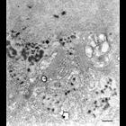
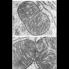
Figure 232 from Chapter 7 (Mitochondria) of 'The Cell, 2nd Ed.' by Don W. Fawcett M.D. Dividing mitochondria in gasric mucosa of a mole. Upper panel, the bilaminar septum as formed, but the outer me...
CIL:10950
NCBI Organism Classification
Felis catus
Biological Process
plasma membrane organization
Cellular Component
basement membrane
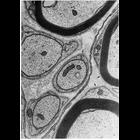
Schwann cells in peripheral nerves secrete a layer similar to the basal lamina called the lamina externa, boundary layer, or basement membrane. In this micrograph, the lamina externa surrounds the Sc...
CIL:39452
NCBI Organism Classification
Vorticella convallaria
Biological Process
micronucleus organization
Cellular Component
micronucleus
Like other ciliates two types of nuclei are present. A germinative micronucleus and a RNA-producing macronucleus. Both have nuclear envelopes with pores and both have ribosomes attached to their outer...
CIL:39791
NCBI Organism Classification
Lytechinus pictus
Biological Process
embryonic morphogenesis
Cellular Component
extracellular matrix
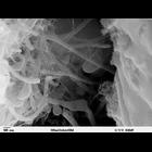
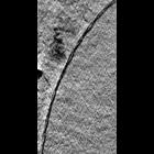
Scanning electron microscope image of Strongylocentrotus drobachiensus [sea urchin] embryo at the late gastrula stage. This high magnification image of the embryo shows the primary mesenchyme syncytia...
CIL:41360
NCBI Organism Classification
Mus musculus
Biological Process
nuclear envelope organization
Cellular Component
nucleus
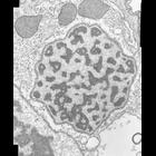
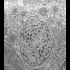
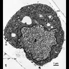
The image shows a slice through a 3D tomographic reconstruction of a mouse adenocarcinoma cell based on x-ray microscopy. Live cells were cryofixed by plunge freezing in liquid ethane and the vitrifi...
CIL:16312
NCBI Organism Classification
Maize mosaic virus
Biological Process
viral reproduction
Cellular Component
host cell nuclear membrane
The images in this group show electron micrographs of epidermal and mesophyll cells in thin sections of maize leaves infected with Maize mosaic virus (MMV, Rhabdoviridae). This jpg image is of an epid...
« Previous
1
2
3
4
5
6
7
8
9
...
1482
Next »
Results per page:
10
20
50
100