Alternate header for print version
Contributors
Help
Submit
Search
menu
Data sets
Videos
Latest data
Center for Research in Biological Systems
Basic Science Building, Room 1000
University of California, San Diego
9500 Gilman Drive
La Jolla, CA 92093-0608, USA
Voice
: (858) 534-0276
Fax
: (858) 534-7497
Email
: dorloff@ncmir.ucsd.edu
Search Results for
Outer nuclear layer of retina
(14816 results)
CIL:11817
NCBI Organism Classification
Danio rerio
Biological Process
anatomical structure morphogenesis
Cellular Component
nucleus
4D data set of yolk syncytial layer nuclear movements. Movie shows z-series stacks sequentially through time. Numbers label individual nuclei so they can be tracked in 3D space at each time point. Vi...
CCDB:8495
Species
Mouse
Organ
eye
Cell type
retina rod cell
System
central nervous system
Structure
rod spherule
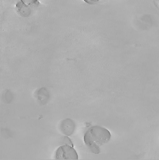
Cone and rod mitochondria: electron tomography
CIL:40386
NCBI Organism Classification
Psidium guajava
Biological Process
plant stem organization
Cellular Component
mucilage
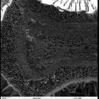
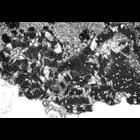
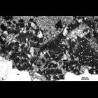
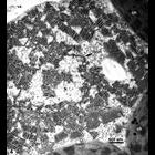
Scanning electron microscope image of cross-section through a Psidium guajava stem. The image shows the thin outer epidermal layer with a thick layer of cortex beneath. The vascular area consists of ...
CIL:36262
NCBI Organism Classification
Vorticella convallaria
Biological Process
nucleus organization
Cellular Component
nuclear envelope
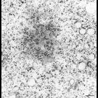
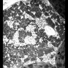
A grazing section through the edge of the nuclear envelope. This illustrates nuclear envelope pores in face view. Polysomes, chains of ribosomes, are bound to the outer membrane of the nuclear envelop...
CIL:12416
NCBI Organism Classification
Maize mosaic virus
Biological Process
viral reproduction
Cellular Component
host cell nuclear membrane
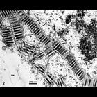
The images in this group show electron micrographs of epidermal and mesophyll cells in thin sections of maize leaves infected with Maize mosaic virus (MMV, Rhabdoviridae). MMV virions are bullet-shape...
CIL:12417
NCBI Organism Classification
Maize mosaic virus
Biological Process
viral reproduction
Cellular Component
host cell nuclear membrane
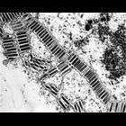
The images in this group show electron micrographs of epidermal and mesophyll cells in thin sections of maize leaves infected with Maize mosaic virus (MMV, Rhabdoviridae). MMV virions are bullet-shape...
CIL:16310
NCBI Organism Classification
Maize mosaic virus
Biological Process
viral reproduction
Cellular Component
host cell nuclear membrane
The images in this group show electron micrographs of epidermal and mesophyll cells in thin sections of maize leaves infected with Maize mosaic virus (MMV, Rhabdoviridae). MMV virions are bullet-shape...
CIL:12415
NCBI Organism Classification
Maize mosaic virus
Biological Process
viral reproduction
Cellular Component
host cell nuclear membrane
The images in this group show electron micrographs of epidermal and mesophyll cells in thin sections of maize leaves infected with Maize mosaic virus (MMV, Rhabdoviridae). MMV virions are bullet-shape...
CIL:16309
NCBI Organism Classification
Maize mosaic virus
Biological Process
viral reproduction
Cellular Component
host cell nuclear membrane
The images in this group show electron micrographs of epidermal and mesophyll cells in thin sections of maize leaves infected with Maize mosaic virus (MMV, Rhabdoviridae). MMV virions are bullet-shape...
CIL:16311
NCBI Organism Classification
Maize mosaic virus
Biological Process
viral reproduction
Cellular Component
host cell nuclear membrane
The images in this group show electron micrographs of epidermal and mesophyll cells in thin sections of maize leaves infected with Maize mosaic virus (MMV, Rhabdoviridae). MMV virions are bullet-shape...
« Previous
1
2
3
4
5
6
7
8
9
...
1482
Next »
Results per page:
10
20
50
100