Alternate header for print version
Contributors
Help
Submit
Search
menu
Data sets
Videos
Latest data
Center for Research in Biological Systems
Basic Science Building, Room 1000
University of California, San Diego
9500 Gilman Drive
La Jolla, CA 92093-0608, USA
Voice
: (858) 534-0276
Fax
: (858) 534-7497
Email
: dorloff@ncmir.ucsd.edu
Search Results for
Secretory granule
(140 results)
CIL:37236
NCBI Organism Classification
Cavia porcellus
Biological Process
protein secretion
Cellular Component
rough endoplasmic reticulum
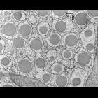
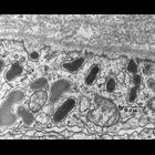
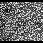
Early transmission electron micrograph of secretory cell from the guinea pig pancreas. Secretory granules are prominent in the lumen of the rough endoplasmic reticulum. Image made available by James ...
CIL:11376
NCBI Organism Classification
Rattus
Biological Process
post-translational protein modification
Cellular Component
Golgi apparatus
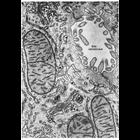
Figure 209 from Chapter 6 (Golgi Apparatus) of 'The Cell, 2nd Ed.' by Don W. Fawcett M.D. Portions of two hepatic cells and the intervening bile canaliculus from rat liver. Two stacks of Golgi ciste...
CIL:7595
NCBI Organism Classification
Rattus
Biological Process
secretion
Cellular Component
secretory granule
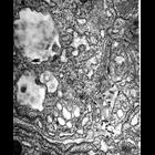
This image shows a group of secretory vesicles loaded with very low density lipoproteins located near the sinusoidal cell membrane of a hepatocyte. The fate of these VLDLs is to be secreted into the b...
CIL:7596
NCBI Organism Classification
Rattus
Biological Process
secretion
Cellular Component
secretory granule
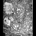
This image shows a group of secretory vesicles loaded with very low density lipoproteins (VLDLs) located near the sinusoidal cell membrane (CM) of a hepatocyte. The fate of these VLDLs is to be secret...
CIL:7649
NCBI Organism Classification
Rattus
Biological Process
exocytosis
Cellular Component
extracellular matrix
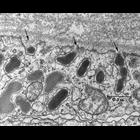
Stimulated Exocytosis. This micrograph shows the peripheral cytoplasm of a mammotroph in the pituitary of a lactating rat. Several secretion granules are lined up facing the perivascular spaces and a...
CIL:9537
NCBI Organism Classification
Rattus
Biological Process
exocytosis
Cellular Component
extracellular matrix
Stimulated Exocytosis. This micrograph shows the peripheral cytoplasm of a mammotroph in the pituitary of a lactating rat. Several secretion granules are lined up facing the perivascular spaces and a...
CIL:10970
NCBI Organism Classification
Rhipicephalus appendiculatus
Biological Process
nucleus organization
Cellular Component
nucleus
Transmission electron micrograph of the salivary gland of the tick Rhipicelphals appendiculatus illustrating the dramatic difference in nuclear size seen in different cell types. Here, the large (prob...
CIL:25378
NCBI Organism Classification
Maize mosaic virus
Biological Process
viral transmission by vector
Cellular Component
vesicle
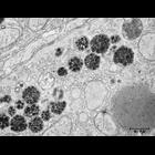
Electron micrograph of Maize mosaic virus (MMV, Rhabdoviridae) in secretory cells from the principal salivary gland of the insect vector Peregrinus maidis (planthopper, Hemiptera, Delphacidae). MMV vi...
CIL:25377
NCBI Organism Classification
Maize mosaic virus
Biological Process
viral transmission by vector
Cellular Component
vesicle
Electron micrograph of Maize mosaic virus (MMV, Rhabdoviridae) in secretory cells from the principal salivary gland of the insect vector Peregrinus maidis (planthopper, Hemiptera, Delphacidae). MMV vi...
CIL:37264
NCBI Organism Classification
Bos taurus
Biological Process
none specified
Cellular Component
zymogen granule
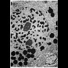
Zymogen granule fraction Calf. Section through a pellet of a zymogen granule fraction showing that the preparation mostly consists of morphologically unaltered zymogen granules. Image made available...
« Previous
1
2
3
4
5
6
7
8
9
...
14
Next »
Results per page:
10
20
50
100