Alternate header for print version
Contributors
Help
Submit
Search
menu
Data sets
Videos
Latest data
Center for Research in Biological Systems
Basic Science Building, Room 1000
University of California, San Diego
9500 Gilman Drive
La Jolla, CA 92093-0608, USA
Voice
: (858) 534-0276
Fax
: (858) 534-7497
Email
: dorloff@ncmir.ucsd.edu
Search Results for
Cortex of kidney
(14820 results)
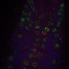
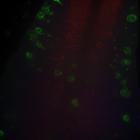
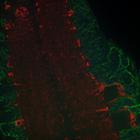
CIL:54649
NCBI Organism Classification
Drosophila melanogaster
Biological Process
Glial-Glial Tiling
Cellular Component
Astrocyte membranes
Confocal image of the CNS of a control D. melanogaster in the third instar larval stage, 76uM from the ventral surface, depicting astrocytes (red), cortex glia (green) and neuronal nuclei (blue). Our ...
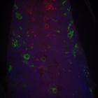
CIL:54647
NCBI Organism Classification
Drosophila melanogaster
Biological Process
Glial-Glial Tiling
Cellular Component
Astrocyte membranes
Confocal image of the CNS of a control D. melanogaster in the third instar larval stage, 62uM from the ventral surface, depicting astrocytes (red), cortex glia (green) and neuronal nuclei (blue). Our ...
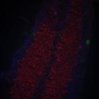
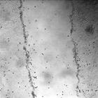
CIL:44506
NCBI Organism Classification
Canis lupus familiaris
Biological Process
wound healing, spreading of epidermal cells
Cellular Component
none specified
Time series light microscopy images illustrating a wound healing assay. A monolayer of MDCK (Madin-Darby Canine Kidney) epithelial cells is scratched to create a 'wound' about 300 micrometers in width...
CIL:44510
NCBI Organism Classification
Canis lupus familiaris
Biological Process
wound healing, spreading of epidermal cells
Cellular Component
none specified
Time series light microscopy images illustrating a wound healing assay. A monolayer of MDCK (Madin-Darby Canine Kidney) epithelial cells is scratched to create a 'wound' about 300 micrometers in width...
CIL:54679
NCBI Organism Classification
Drosophila melanogaster
Biological Process
Glial-Glial Tiling
Cellular Component
Astrocyte membranes
Confocal image of the CNS of a D. melanogaster in the third instar larval stage after αSNAP KD, 14uM from the ventral surface, depicting astrocytes (red), cortex glia (green) and neuronal nuclei (bl...
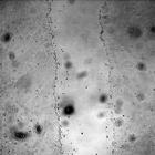
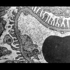
CIL:37181
NCBI Organism Classification
Rattus
Biological Process
glomerular filtration
Cellular Component
basement membrane
Transmission electron micrograph of rat kidney filtration barrier. Image is low magnification vies of the basement membrane filtration slits, urinary space, and the glomerular filtration capillary lu...
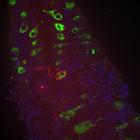
CIL:54678
NCBI Organism Classification
Drosophila melanogaster
Biological Process
Glial-Glial Tiling
Cellular Component
Astrocyte membranes
Confocal image of the CNS of a D. melanogaster in the third instar larval stage after αSNAP KD, 24uM from the ventral surface, depicting astrocytes (red), cortex glia (green) and neuronal nuclei (bl...
CIL:54673
NCBI Organism Classification
Drosophila melanogaster
Biological Process
Glial-Glial Tiling
Cellular Component
Astrocyte membranes
Confocal image of the CNS of a D. melanogaster in the third instar larval stage after αSNAP KD, 28uM from the ventral surface, depicting astrocytes (red), cortex glia (green) and neuronal nuclei (bl...
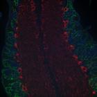
CIL:54675
NCBI Organism Classification
Drosophila melanogaster
Biological Process
Glial-Glial Tiling
Cellular Component
Astrocyte membranes
Confocal image of the CNS of a D. melanogaster in the third instar larval stage after αSNAP KD, 82uM from the ventral surface, depicting astrocytes (red), cortex glia (green) and neuronal nuclei (bl...
CIL:54682
NCBI Organism Classification
Drosophila melanogaster
Biological Process
Glial-Glial Tiling
Cellular Component
Astrocyte membranes
Confocal image of the CNS of a D. melanogaster in the third instar larval stage after αSNAP KD, 8uM from the ventral surface, depicting astrocytes (red), cortex glia (green) and neuronal nuclei (blu...
« Previous
1
2
3
4
5
6
7
8
9
...
1482
Next »
Results per page:
10
20
50
100

")