Alternate header for print version
Contributors
Help
Submit
Search
menu
Data sets
Videos
Latest data
Center for Research in Biological Systems
Basic Science Building, Room 1000
University of California, San Diego
9500 Gilman Drive
La Jolla, CA 92093-0608, USA
Voice
: (858) 534-0276
Fax
: (858) 534-7497
Email
: dorloff@ncmir.ucsd.edu
Search Results for
Cortex of kidney
(14820 results)
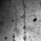
CIL:37175
NCBI Organism Classification
Rattus
Biological Process
glomerular filtration
Cellular Component
none specified
Transmission electron micrograph of kidney nephrotic glomerulus. The series of images in this group have particular historic and cell biologic importance for determining the role of the glomerular b...
CIL:39041
NCBI Organism Classification
Macaca mulatta
Biological Process
pathogenesis
Cellular Component
nucleus
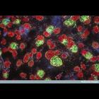
Photomicrograph of Chlamydia grown in culture. The sample was taken from rhesus monkey kidney cells and stained with giemsa. The cell nuclei appear red and the infective 'elementary bodies' of the Chl...
CIL:9973
NCBI Organism Classification
Rattus
Biological Process
constitutive secretory pathway
Cellular Component
Golgi apparatus
Normal Rat Kidney (NRK) cells grown in culture expressing Galactosyl Transferase-YFP (GalT-YFP) and p58-CFP. This file is the YFP time series demonstrating the organization of the early secretory path...
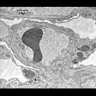
CIL:9535
NCBI Organism Classification
Rattus
Biological Process
none specified
Cellular Component
basement membrane
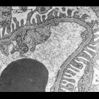
Peripheral area of normal rat kidney. The capillary wall is composed of three distinct layers: the endothelium with its fenestra, the basement membrane (BM) which is a continuous layer 0.1 to 0.15 mic...
CIL:37179
NCBI Organism Classification
Rattus
Biological Process
glomerular filtration
Cellular Component
none specified
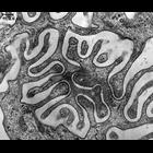
Transmission electron micrograph of rat kidney interdigitating foot processes at high magnification. The series of images in this group have particular historic and cell biologic importance for dete...
CIL:44502
NCBI Organism Classification
Canis lupus familiaris
Biological Process
wound healing, spreading of epidermal cells
Cellular Component
none specified
Time series light microscopy images illustrating a wound healing assay. A monolayer of MDCK (Madin-Darby Canine Kidney) epithelial cells is scratched to create a 'wound' about 300 micrometers in width...
CIL:44507
NCBI Organism Classification
Canis lupus familiaris
Biological Process
wound healing, spreading of epidermal cells
Cellular Component
none specified
Time series light microscopy images illustrating a wound healing assay. A monolayer of MDCK (Madin-Darby Canine Kidney) epithelial cells is scratched to create a 'wound' about 300 micrometers in width...
CIL:44511
NCBI Organism Classification
Canis lupus familiaris
Biological Process
wound healing, spreading of epidermal cells
Cellular Component
none specified
Movie created from time series light microscopy images (CIL:44501) illustrating a wound healing assay. A monolayer of MDCK (Madin-Darby Canine Kidney) epithelial cells is scratched to create a 'wound'...
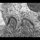
CIL:37168
NCBI Organism Classification
Rattus
Biological Process
adherens junction organization
Cellular Component
adherens junction
Transmission electron micrograph of adherens junctions (macula adherens or desmosome)in the proximal tubule of a rat kidney. The junctions with associated tonofilaments are seen at the apposed plasma...
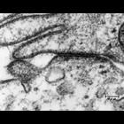
CIL:37172
NCBI Organism Classification
none specified
Biological Process
tight junction assembly
Cellular Component
tight junction
Transmission electron micrograph of tight junction (zonula occludens) between adjacent plasma membranes in the distal kidney tubule. The darkly staining horizontal structure forms the junctional compl...
« Previous
1
2
3
4
5
6
7
8
9
...
1482
Next »
Results per page:
10
20
50
100

")