Alternate header for print version
Contributors
Help
Submit
Search
menu
Data sets
Videos
Latest data
Center for Research in Biological Systems
Basic Science Building, Room 1000
University of California, San Diego
9500 Gilman Drive
La Jolla, CA 92093-0608, USA
Voice
: (858) 534-0276
Fax
: (858) 534-7497
Email
: dorloff@ncmir.ucsd.edu
Search Results for
nucleus organization
(261 results)
CIL:36646
NCBI Organism Classification
Paramecium multimicronucleatum
Biological Process
macronucleus organization
Cellular Component
macronucleus
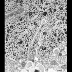
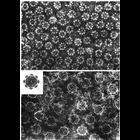
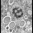
The macronucleus is the largest organelle in the P. multimicronucleatum cell and can measure 35µm long by 12µm wide. A small fraction of a macronucleus is seen in this micrograph. The chromatin appe...
CIL:34536
NCBI Organism Classification
Tetrahymena pyriformis
Biological Process
macronucleus organization
Cellular Component
macronucleus
High resolution view of the nuclear envelope of both the macronucleus, seen here, and the micronucleus consists of 2 membranes containing numerous perforations called pores. Proteins are imported thro...
CIL:41360
NCBI Organism Classification
Mus musculus
Biological Process
nuclear envelope organization
Cellular Component
nucleus
The image shows a slice through a 3D tomographic reconstruction of a mouse adenocarcinoma cell based on x-ray microscopy. Live cells were cryofixed by plunge freezing in liquid ethane and the vitrifi...
CIL:41363
NCBI Organism Classification
Mus musculus
Biological Process
nucleus organization
Cellular Component
mitochondrion
The image shows a slice through a 3D tomographic reconstruction of a mouse adenocarcinoma cell based on x-ray microscopy. Live cells were cryofixed by plunge freezing in liquid ethane and the vitrifi...
CIL:41364
NCBI Organism Classification
Mus musculus
Biological Process
nuclear envelope organization
Cellular Component
nucleus
The image shows a slice through a 3D tomographic reconstruction of a mouse adenocarcinoma cell based on x-ray microscopy. Live cells were cryofixed by plunge freezing in liquid ethane and the vitrifi...
CIL:8101
NCBI Organism Classification
Drosophila melanogaster
Biological Process
mitotic nuclear envelope disassembly
Cellular Component
nucleus
This Drosophila melanogaster early embryo (cycles 10-12) was injected with a fluorescent protein containing a nuclear localization signal (GFP-NLS). Proteins that contain a NLS are recognized by impor...
CIL:11052
NCBI Organism Classification
Taricha granulosa
Biological Process
nucleus organization
Cellular Component
nuclear envelope
Early transmission electron micrographs showing pore details in negatively stained preparations of nuclear envelopes. Upper panel shows the pronounced 8-fold symmetry in the closely spaced pores of t...
CIL:15798
NCBI Organism Classification
Dendraster excentricus
Biological Process
mitosis
Cellular Component
nucleus
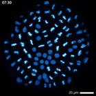
Time lapse movie of a developing sand dollar embryo expressing GFP-histone H2B. The movie shows several rounds of mitoses.
CIL:36645
NCBI Organism Classification
Paramecium multimicronucleatum
Biological Process
micronucleus organization
Cellular Component
micronucleus
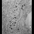
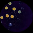
Like other ciliates P. multimicronucleatum has two morphologically and functionally different nuclei. Shown here is the tiny micronucleus, of which there are four, during interphase of this cell. This...
CIL:36648
NCBI Organism Classification
Paramecium multimicronucleatum
Biological Process
nuclear pore distribution
Cellular Component
nuclear pore
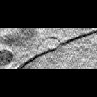
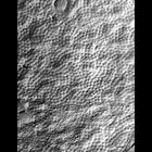
A freeze-fractured view of the nuclear envelope. The fracture in the middle of the figure exposes the cytosolic facing membrane. This can be distinguished by a fractured cytoplasmic organelle at the t...
« Previous
1
2
3
4
5
6
7
8
9
...
27
Next »
Results per page:
10
20
50
100