Alternate header for print version
Contributors
Help
Submit
Search
menu
Data sets
Videos
Latest data
Center for Research in Biological Systems
Basic Science Building, Room 1000
University of California, San Diego
9500 Gilman Drive
La Jolla, CA 92093-0608, USA
Voice
: (858) 534-0276
Fax
: (858) 534-7497
Email
: dorloff@ncmir.ucsd.edu
Search Results for
nucleus organization
(261 results)
CIL:10977
NCBI Organism Classification
Cavia porcellus
Biological Process
nucleus organization
Cellular Component
nuclear heterochromatin
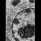
Transmission electron micrograph showing the extensive accumulation of darkly stained heterochromatin at the nuclear periphery typical of many differentiated cells. Heterochromatin is excluded from re...
CIL:11027
NCBI Organism Classification
Didelphimorphia
Biological Process
nucleus organization
Cellular Component
nucleolus
Transmission electron micrographs of Opossum spermatogonia showing different regions within the nucleolus. One region is more lightly staining and fine textured, the other denser and more coarsely tex...
CIL:11031
NCBI Organism Classification
Batrachoseps attenuatus
Biological Process
nucleus organization
Cellular Component
nucleolus
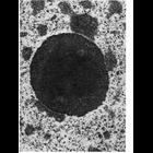
Transmission electron micrograph showing the highly compact nucleolus in the salamander hepatocyte. A finer textured central region is surrounded by a coarser region.
CIL:40315
NCBI Organism Classification
Homo sapiens
Biological Process
nucleus organization
Cellular Component
nuclear pore
Transmission electron micrograph of a thin section of the nucleus of a HeLa cell expressing mutant ALADIN. Pore structure appears to be unaffected in the mutant. From a study of the role of the nuclea...
CIL:10987
NCBI Organism Classification
Unspecified
Biological Process
nucleus organization
Cellular Component
nucleus
Transmission electron micrograph of glutaraldehyde fixed pancreatic acinar cell showing the characteristic features of nuclear chromatin with this preparative method. Darkly staining heterochromatin (...
CIL:11054
NCBI Organism Classification
Anura
Biological Process
nucleus organization
Cellular Component
nuclear envelope
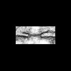
Transmission electron micrographs of sections of tadpole oocyte show the potential for exchange between the nucleus and the cytoplasm at the nuclear envelope. Thin filaments that appear to traverse nu...
CIL:40314
NCBI Organism Classification
Homo sapiens
Biological Process
nucleus organization
Cellular Component
nuclear pore
Transmission electron micrograph of a thin section of the nucleus of a normal HeLa cell showing detail of a cross section of a nuclear pore - from a study of the role of the nuclear porin ALADIN. Oth...
CIL:40317
NCBI Organism Classification
Homo sapiens
Biological Process
nucleus organization
Cellular Component
nuclear pore
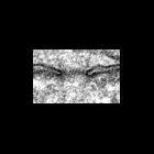
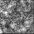
Transmission electron micrograph of a thin section of the nucleus of a normal HeLa cell showing a tangential view of the nuclear envelope with nuclear pores. From a study of the role of the nuclear po...
CIL:11048
NCBI Organism Classification
Cavia porcellus
Biological Process
nucleus organization
Cellular Component
nuclear envelope
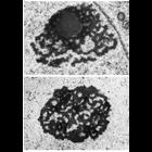
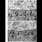
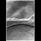
Nuclear pores are clearly seen in these transmission electron micrographs of cells after cryo-fixation, freeze-fracture and surface replication. Upper panel shows the outer surface of of the nuclear e...
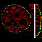
CIL:40313
NCBI Organism Classification
Homo sapiens
Biological Process
nucleus organization
Cellular Component
nucleus
Fluorescence images showing the distribution of GFP-ALADIN, a WD-repeat protein that is mutated in the human triple A syndrome (green), and Tpr, a nucleoporin that localizes to the nucleoplasmic filam...
« Previous
1
2
3
4
5
6
7
8
9
...
27
Next »
Results per page:
10
20
50
100