Alternate header for print version
Contributors
Help
Submit
Search
menu
Data sets
Videos
Latest data
Center for Research in Biological Systems
Basic Science Building, Room 1000
University of California, San Diego
9500 Gilman Drive
La Jolla, CA 92093-0608, USA
Voice
: (858) 534-0276
Fax
: (858) 534-7497
Email
: dorloff@ncmir.ucsd.edu
Search Results for
endocytosis
(99 results)
CIL:11150
NCBI Organism Classification
Homo sapiens
Biological Process
pinocytosis
Cellular Component
coated vesicle
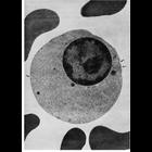
This transmission electron micrograph of a section through a late orthochromatic erythroblast (normoblast) from human bone marrow shows shallow depressions along the membrane surface, indicated by arr...
CIL:13002
NCBI Organism Classification
none specified
Biological Process
pinocytosis
Cellular Component
plasma membrane
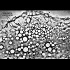
Phase contrast microscopy of living cells offers the ability to observe dynamic behaviors. This single frame image shows folds in the membrane at the periphery of a cell in growing in vitro. Asteris...
CIL:10276
NCBI Organism Classification
Mus musculus
Biological Process
platelet-derived growth factor receptor signaling pathway
Cellular Component
dynamin
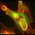
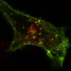
After over-night serum starvation, mouse fibroblast expressing Dyn2-GFP were stimulated with 10ng/ml PDGF. After 5min, the cells were fixed and stained with rhodamine-phalloidin. PDGF-induced dorsal m...
CIL:11127
NCBI Organism Classification
none specified
Biological Process
pinocytosis
Cellular Component
plasma membrane
Phase contrast microscopy of living cells offers the ability to observe dynamic behaviors. This single frame image shows folds in the membrane at the periphery of a cell in growing in vitro. Image b...
CIL:31940
NCBI Organism Classification
Homo sapiens
Biological Process
G-protein coupled receptor signaling pathway
Cellular Component
integral to plasma membrane
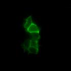
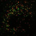
Time-lapse experiments showing deltorphin II (100 nM; a selective agonist)-induced internalization of delta-opioid receptor–enhanced GFP (DOR-EGFP; green) receptors expressed in HEK 293 cells. The C...
CIL:13700
NCBI Organism Classification
Chlorocebus aethiops
Biological Process
G-protein coupled receptor internalization
Cellular Component
plasma membrane
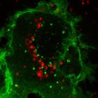
This is one of a group of four images in Figure S5 in Apaja et al., JCB 2010, that support the conclusion that the WT vasopressin type2 receptor (V2R) and the V2R W164S mutant (that causes nephrogenic...
CIL:13699
NCBI Organism Classification
Chlorocebus aethiops
Biological Process
G-protein coupled receptor internalization
Cellular Component
plasma membrane
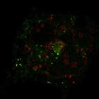
This is one of a group of four images in Figure S5 in Apaja et al., JCB 2010, that support the conclusion that the WT vasopressin type2 receptor (V2R) and the V2R W164S mutant (that causes nephrogenic...
CIL:13701
NCBI Organism Classification
Chlorocebus aethiops
Biological Process
G-protein coupled receptor internalization
Cellular Component
plasma membrane
This is one of a group of four images in Figure S5 in Apaja et al., JCB 2010, that support the conclusion that the WT vasopressin type2 receptor (V2R) and the V2R W164S mutant (that causes nephrogenic...
CIL:13698
NCBI Organism Classification
Chlorocebus aethiops
Biological Process
G-protein coupled receptor internalization
Cellular Component
plasma membrane
This is one of a group of four images in Figure S5 in Apaja et al., JCB 2010, that support the conclusion that the WT vasopressin type2 receptor (V2R) and the V2R W164S mutant (that causes nephrogenic...
CIL:10850
NCBI Organism Classification
Chinchilla
Biological Process
phagocytosis
Cellular Component
lysosome
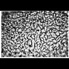
Figure 275 from Chapter 8 (Lysosomes) of 'The Cell, 2nd Ed.' by Don W. Fawcett M.D. Lysosomes of cells engaged in heterophagy, like the Sertoli cells from the testis of the chinchilla shown here, are...
« Previous
1
2
3
4
5
6
7
8
9
10
Next »
Results per page:
10
20
50
100