Alternate header for print version
Contributors
Help
Submit
Search
menu
Data sets
Videos
Latest data
Center for Research in Biological Systems
Basic Science Building, Room 1000
University of California, San Diego
9500 Gilman Drive
La Jolla, CA 92093-0608, USA
Voice
: (858) 534-0276
Fax
: (858) 534-7497
Email
: dorloff@ncmir.ucsd.edu
Search Results for
cellular membrane organization
(334 results)
CIL:36648
NCBI Organism Classification
Paramecium multimicronucleatum
Biological Process
nuclear pore distribution
Cellular Component
nuclear pore
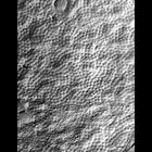
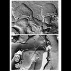
A freeze-fractured view of the nuclear envelope. The fracture in the middle of the figure exposes the cytosolic facing membrane. This can be distinguished by a fractured cytoplasmic organelle at the t...
CIL:17892
NCBI Organism Classification
Didinium nasutum
Biological Process
phagocytosis
Cellular Component
oral apparatus
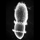
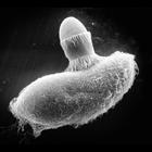
Didinium ingests Paramecium. Also showing a few discharged trichocysts and metachronous waves of cilia in the two characteristic ciliary girdles of Didinium nasutum. This micrograph was taken in 1968 ...
CIL:19537
NCBI Organism Classification
Didinium nasutum
Biological Process
oral apparatus organization
Cellular Component
oral apparatus
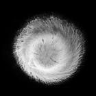
The proboscis of Didinium. While not well preserved, this image clearly shows the clockwise surface ridges that are elevated by ribbons of microtubules that are involved in supporting the buccal cavit...
CIL:21991
NCBI Organism Classification
Didinium nasutum
Biological Process
phagocytosis
Cellular Component
oral apparatus
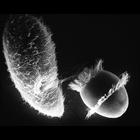
Didinium captures Paramecium. A moment after initial contact when toxicysts enter Paramecium. The strand of toxicysts can be seen between the two organisms, and Didinium will use the anchored toxicyst...
CIL:39250
NCBI Organism Classification
Didinium nasutum
Biological Process
phagocytosis
Cellular Component
oral apparatus
Didinium captures Paramecium. It is not unusual for an undersized Didinium to attempt to ingest a much larger Paramecium. In this case, the organisms were preserved before the outcome could be determi...
CIL:10914
NCBI Organism Classification
Macaca mulatta
Biological Process
neurotransmitter secretion
Cellular Component
synapse part
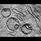
Freeze fracture images of synaptic junctions in the retina of Macaca mulatta. The electron micrograph in the top panel shows two invaginating synapses between cone and horizontal cells in the outer p...
CIL:41359
NCBI Organism Classification
Mus musculus
Biological Process
cytoplasm organization
Cellular Component
mitochondrion
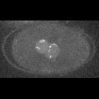
The image shows a portion of a slice through a 3D tomographic reconstruction of a mouse adenocarcinoma cell based on x-ray microscopy. Live cells were cryofixed by plunge freezing in liquid ethane an...
CIL:24922
NCBI Organism Classification
uncultured Scuticociliatia
Biological Process
cortical cytoskeleton organization
Cellular Component
macronucleus
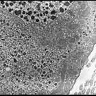
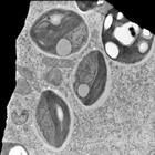
Ancistrum is a marine ciliate that inhabits the mantle cavity of mytiliid mussels. This image shows a good view of the macronucleus, mitochondria with tubular cristae, and what are probably flask-shap...
CIL:28783
NCBI Organism Classification
Caenorhabditis elegans
Biological Process
KNL-1 depeletion
Cellular Component
nuclear chromosome
Spinning disk confocal time-lapse imaging of the first mitosis in a C. elegans embryo expressing GFP-KNL-2 (strain OD31) in which KNL-1 has been depleted by RNAi. Discrete foci of GFP signal within th...
CIL:25697
NCBI Organism Classification
Trichodina
Biological Process
response to symbiont
Cellular Component
symbiont-containing vacuole
A view of the endosymbiotic Chlorella of a green Trichodina that lives on the surface of a freshwater snail collected in central Illinois. In this image there is also an endosymbiotic bacterium. It ha...
« Previous
1
...
3
4
5
6
7
8
9
10
...
34
Next »
Results per page:
10
20
50
100