Alternate header for print version
Contributors
Help
Submit
Search
menu
Data sets
Videos
Latest data
Center for Research in Biological Systems
Basic Science Building, Room 1000
University of California, San Diego
9500 Gilman Drive
La Jolla, CA 92093-0608, USA
Voice
: (858) 534-0276
Fax
: (858) 534-7497
Email
: dorloff@ncmir.ucsd.edu
Search Results for
cell-cell junction organization
(43 results)
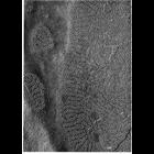
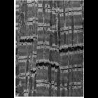
CIL:11204
NCBI Organism Classification
Siphonaptera
Biological Process
cell-cell junction organization
Cellular Component
cell-cell junction
Freeze-fracture preparation of a continuous junction on the lateral surface of epithelial cells from the gut of a flea. Meandering ridges appear to be on the P-face of the replica. Figure 80, by Susu...
CIL:11226
NCBI Organism Classification
Mus musculus
Biological Process
cell communication
Cellular Component
gap junction
Gap junctions isolated from mouse liver and negatively stained with uranyl formate. At higher magnification, the 8-9 nm particles, or connexons, have a central 1.5-2 nm electron dense pore. Images b...
CIL:11230
NCBI Organism Classification
Rattus
Biological Process
cell communication
Cellular Component
gap junction
Gap junctions from a granulosa cell of a rat ovarian follicle. Variability in size of the gap junctions is likely an artifact of the freeze fracture preparation. Figure 99 from Chapter 3 (Junctional ...
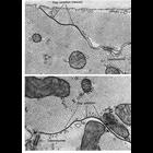
CIL:11232
NCBI Organism Classification
none specified
Biological Process
cell communication
Cellular Component
gap junction
Gap junctions in ciliary epithelium under conditions of oxygenation (upper) and anoxia (lower), in tissue prepared by ultrarapid freezing using liquid helium. Under oxygenated conditions, the connexo...
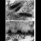
CIL:11243
NCBI Organism Classification
Felis catus
Biological Process
cell communication
Cellular Component
gap junction
Papillary muscle from cat heart shows a step-like end-to-end junction of two cardiac muscle cells. The longitudinally oriented lateral cell boundaries are straight and exhibit extensive gap junctions...
CIL:11245
NCBI Organism Classification
Felis catus
Biological Process
cell communication
Cellular Component
gap junction
Gap junctions in papillary muscle from cat heart. These communicating junctions are responsible for the spreading wave of depolarization through the myocardium. Desmosomes can also be found on the l...
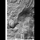
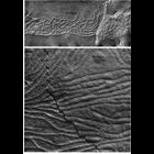
CIL:11209
NCBI Organism Classification
Phodopus
Biological Process
cell adhesion
Cellular Component
desmosome
Upper panel, adjoining portions of two cells in the stratum spinosum of hamster cheek pouch epithelium shows desmosome junctions between two cells. Lower, a portion of the basal surface of a cell in ...
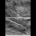
CIL:37185
NCBI Organism Classification
Rattus
Biological Process
cell-cell junction organization
Cellular Component
cell-cell junction
Transmission electron micrograph of the junctional complexes at the intersection of three hepatocytes. This image was taken from liver tissues from an ethanol fed rat and also shows the bile canalic...
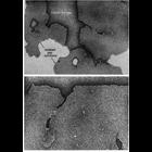
CIL:11196
NCBI Organism Classification
Rattus
Biological Process
cell-cell junction organization
Cellular Component
occluding junction
Upper panel shows a typical zonula occludens of rat intestinal epithelium, prepared using freeze fracture techniques. A network of intersecting strands on the P-face, and a complementary pattern of g...
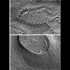
CIL:11237
NCBI Organism Classification
Macaca mulatta
Biological Process
cell communication
Cellular Component
gap junction
Representative examples of gap junctions from vertebrates (ciliary epithelium from the eye of a Macaca mulatta, upper) and invertebrates (inverted gap junctions from cells of the mid-gut of the horses...
« Previous
1
2
3
4
5
Next »
Results per page:
10
20
50
100