Alternate header for print version
Contributors
Help
Submit
Search
menu
Data sets
Videos
Latest data
Center for Research in Biological Systems
Basic Science Building, Room 1000
University of California, San Diego
9500 Gilman Drive
La Jolla, CA 92093-0608, USA
Voice
: (858) 534-0276
Fax
: (858) 534-7497
Email
: dorloff@ncmir.ucsd.edu
Search Results for
cell migration
(92 results)
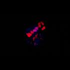
CIL:11806
NCBI Organism Classification
Danio rerio
Biological Process
cell migration
Cellular Component
nucleus
Patterned nuclei movements in the dorsal yolk syncytial layer( YSL) after mid-gastrulation. Anterior is up. Timelapse includes Nomarski images (left panel), fluorescence (right panel) and overlay (mid...
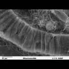
CIL:39786
NCBI Organism Classification
Lytechinus pictus
Biological Process
embryonic morphogenesis
Cellular Component
cilium
Scanning electron microscope image of Strongylocentrotus drobachiensus [sea urchin] at the gastrula stage. Embryo was split open to reveal a nice cross-section through the outer epithelial layer and t...
CIL:11822
NCBI Organism Classification
Danio rerio
Biological Process
anatomical structure morphogenesis
Cellular Component
nucleus
Yolk syncytial layer cell nuclei exhibit convergence and extension behaviors during late gastrulation. Video made available by Mark Cooper through Zebrafish - The Living Laboratory.
CIL:25539
NCBI Organism Classification
Drosophila melanogaster
Biological Process
heart development
Cellular Component
nucleus
A 3D reconstruction of the cardiac outflow tract (OFT) region (horizontal and vertical rotation) from stage-16 wild-type embryo stained for Ladybird early (Lbe; blue) and Tinman (Tin; red). Heart-anch...
CIL:25538
NCBI Organism Classification
Drosophila melanogaster
Biological Process
cardiac cell differentiation
Cellular Component
nucleus
A 3D reconstruction of the cardiac outflow tract (OFT) region (horizontal and vertical rotation) from stage-14 wild-type embryo stained for Tinman (Tin; red) and Msp300 (green). Tin staining reveals t...
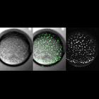
CIL:39049
NCBI Organism Classification
none specified
Biological Process
substrate-dependent cell migration, cell attachment to substrate
Cellular Component
cell surface
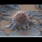
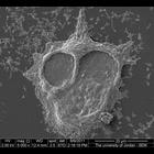
Colorized scanning electron micrograph of a lung cancer cell grown in culture.
CIL:38974
NCBI Organism Classification
none specified
Biological Process
neuron migration
Cellular Component
growth cone
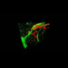
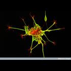
Dorsal root ganglion nerve cells stained to reveal the microtubules (green) and actin filaments (red). The axon shaft contains bundles of microtubules that give structural support and carry cargo (pro...
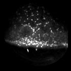
CIL:34864
NCBI Organism Classification
Homo sapiens
Biological Process
leukocyte chemotaxis involved in inflammatory response
Cellular Component
nucleus
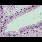
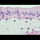
Gallbladder wall of a human patient with gallstones. The duct (clear zone at center) is lined by highly polarized epithelial cells with dark nuclei at the base. At the top of the picture, below the...
CIL:34859
NCBI Organism Classification
Homo sapiens
Biological Process
leukocyte chemotaxis involved in inflammatory response
Cellular Component
nucleus
Gallbladder wall of a human patient with gallstones. The top layer is composed of highly polarized epithelial cells with microvilli partially visible at the apex and dark nuclei at the base. Below ...
CIL:40624
NCBI Organism Classification
Homo sapiens
Biological Process
substrate-dependent cell migration
Cellular Component
cell surface
Scanning electron micrograph of breast cancer cells that have aggregated.
« Previous
1
2
3
4
5
6
7
8
9
10
Next »
Results per page:
10
20
50
100