Alternate header for print version
Contributors
Help
Submit
Search
menu
Data sets
Videos
Latest data
Center for Research in Biological Systems
Basic Science Building, Room 1000
University of California, San Diego
9500 Gilman Drive
La Jolla, CA 92093-0608, USA
Voice
: (858) 534-0276
Fax
: (858) 534-7497
Email
: dorloff@ncmir.ucsd.edu
Search Results for
cell adhesion
(348 results)
CIL:11199
NCBI Organism Classification
Rattus
Biological Process
cell-cell adhesion
Cellular Component
zonula adherens
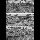
These examples of ependymal epithelium from the rat brain represent epithelia that lack zonulae occludentes. Further, there appears to be no consistent order to the junctional specializations that are...
CIL:40604
NCBI Organism Classification
Amaryllis
Biological Process
pollen adhesion
Cellular Component
pollen tube
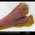
Colorized scanning electron micrograph of Amarylis stigma (pink) with pollen grains (yellow) adhering to sticky glands on its surface. Some pollen tubes (olive green) carrying genetic material can be ...
CIL:41307
NCBI Organism Classification
Penta lanceolata
Biological Process
pollen adhesion
Cellular Component
pollen coat
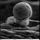
Scanning electron micrograph showing Penta lanceolata unacetolyzed pollen. Compare this pollen to acetolyzed pollen images by the same contributor in the Library. This image is a ]higher magnificatio...
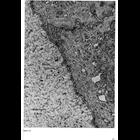
CIL:10948
NCBI Organism Classification
Rattus
Biological Process
cell-substrate adhesion
Cellular Component
basement membrane
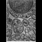
Electron micrograph of a nerve from the mesentery of a rat shows groups of unmyelinated axons wrapped by deeply invaginating Schwann cells in cross section. Surrounding the Schwann cell wrapping is a ...
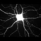
CIL:807
NCBI Organism Classification
Rattus
Biological Process
calcium-dependent cell-cell adhesion
Cellular Component
excitatory synapse
During hippocampal neuron development in vitro, N-cadherin becomes selectively localized to excitatory synapses. This image shows immunolocalization of N-cadherin. Other images in this image group sh...
CIL:10942
NCBI Organism Classification
Homo sapiens
Biological Process
cell-substrate adhesion
Cellular Component
basal lamina
This electron micrograph shows the basal lamina as a thin gray moustache following parallel along the basal membrane of epithelial cells from human corneal epithelium. In this tissue, the basal lamin...
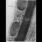
CIL:10944
NCBI Organism Classification
Homo sapiens
Biological Process
cell-substrate adhesion
Cellular Component
basal lamina
This electron micrograph shows the endothelium and the underlying Descemet's membrane of the human cornea. Descemet's membrane is an unusually thick and structurally specialized example of basal lami...
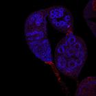
CIL:13741
NCBI Organism Classification
Drosophila melanogaster
Biological Process
smoothened signaling pathway
Cellular Component
P granule
Boi (red) localizes predominantly to apical cells in Drosophila wild-type germaria but not in germaria with UAS-boi[RNAi] expression (CIL#13742). Germ cells are identified using anti-Vasa (blue). Imag...
CIL:13160
NCBI Organism Classification
Homo sapiens
Biological Process
cell migration
Cellular Component
cell
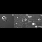
Topographical regulation of keratinocyte migration. 2D, 3D, and 1D fibrillar migration of human epidermal keratinocytes. 2D matrices were constructed by uniform coating with extracellular matrix (ECM)...
CIL:11197
NCBI Organism Classification
Rattus
Biological Process
cell-cell adhesion
Cellular Component
occluding junction
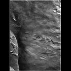
A freeze fracture replica of a Sertoli cell junction from rat testis shows more than 50 rows of particles along the E-face. Image from Gilula, Fawcett and Aoki, Dev. Biol. 50: 142-168 (1976), reprint...
« Previous
1
2
3
4
5
6
7
8
9
...
35
Next »
Results per page:
10
20
50
100