Alternate header for print version
Contributors
Help
Submit
Search
menu
Data sets
Videos
Latest data
Center for Research in Biological Systems
Basic Science Building, Room 1000
University of California, San Diego
9500 Gilman Drive
La Jolla, CA 92093-0608, USA
Voice
: (858) 534-0276
Fax
: (858) 534-7497
Email
: dorloff@ncmir.ucsd.edu
Search Results for
cell adhesion
(348 results)
CIL:38991
NCBI Organism Classification
none specified
Biological Process
epithelial cell-cell adhesion
Cellular Component
cell-cell junction
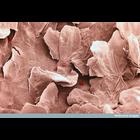
Colorized scanning electron micrograph of keratinised squamous epithelial lining of the upper vagina. The boundaries between adjacent cells are visible as raised ridges. The cells closest to the surfa...
CIL:39085
NCBI Organism Classification
Sisymbrium officinale
Biological Process
pollen adhesion
Cellular Component
cell surface
Colorized scanning electron micrograph of pollen on the anther of a hedge mustard (Sisymbrium officinale) flower. This plant is reputed to have various medicinal properties including its ability to tr...
CIL:7439
NCBI Organism Classification
Mus musculus
Biological Process
cell adhesion
Cellular Component
focal adhesion
Mouse Embryonic Fibroblasts (MEFs) grown on glass coverslips coated with 10 ug/ml Fibronectin. After 24 hrs cells were fixed in 3% paraformaldehyde and stained with mouse antibody to Paxillin, rabbit...
CIL:38957
NCBI Organism Classification
Homo sapiens
Biological Process
receiving nourishment
Cellular Component
zona pellucida
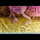
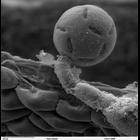
A colorized scanning electron micrograph of a human egg, which is the huge cell colored yellow at the bottom of this image. The follicle cells that surround it (top) send out long projections that pen...
CIL:41309
NCBI Organism Classification
Penta lanceolata
Biological Process
pollen adhesion
Cellular Component
pollen coat
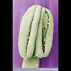
Scanning electron micrograph showing Penta lanceolata unacetolyzed pollen. This is a higher magnification view of CIL 41306. Compare this pollen to acetolyzed pollen images by the same contributor in...
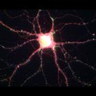
CIL:809
NCBI Organism Classification
Rattus
Biological Process
calcium-dependent cell-cell adhesion
Cellular Component
excitatory synapse
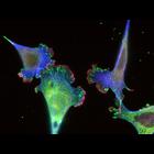
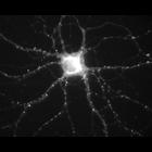
During hippocampal neuron development in vitro, N-cadherin becomes selectively localized to excitatory synapses. This image shows synapses that are also immunopositive for the presynaptic glutamate tr...
CIL:38599
NCBI Organism Classification
Homo sapiens
Biological Process
focal adhesion assembly
Cellular Component
focal adhesion
This total internal reflection (TIRF) image of both the Dronpa-alphaa ctinin and the unconverted tdEos-vinculin channels corresponds to the same image field as the diffraction limited DIC image CIL 38...
CIL:38651
NCBI Organism Classification
Homo sapiens
Biological Process
molecular organization
Cellular Component
focal adhesion
This photoactivation localization microscopy (PALM) image of tdEos-vinculin (red) and Dronpa-paxillin (green) illustrates that vinculin and paxillin are segregated into interlocking microdomains withi...
CIL:808
NCBI Organism Classification
Rattus
Biological Process
calcium-dependent cell-cell adhesion
Cellular Component
excitatory synapse
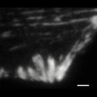
During hippocampal neuron development in vitro, N-cadherin becomes selectively localized to excitatory synapses. This image shows immunolocalization of the NMDA receptor, which is postsynaptic. Other...
CIL:810
NCBI Organism Classification
Rattus
Biological Process
calcium-dependent cell-cell adhesion
Cellular Component
excitatory synapse
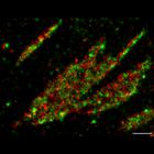
During hippocampal neuron development in vitro, N-cadherin becomes selectively localized to excitatory synapses. This image shows immunolocalization of N-cadherin (red), colocalized at synapses that a...
« Previous
1
2
3
4
5
6
7
8
9
...
35
Next »
Results per page:
10
20
50
100