Alternate header for print version
Contributors
Help
Submit
Search
menu
Data sets
Videos
Latest data
Center for Research in Biological Systems
Basic Science Building, Room 1000
University of California, San Diego
9500 Gilman Drive
La Jolla, CA 92093-0608, USA
Voice
: (858) 534-0276
Fax
: (858) 534-7497
Email
: dorloff@ncmir.ucsd.edu
Search Results for
cell adhesion
(348 results)
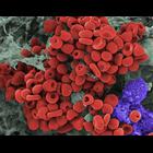
CIL:18042
NCBI Organism Classification
Mesocricetus auratus
Biological Process
erythrocyte aggregation
Cellular Component
cell
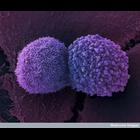
Scanning electron micrograph of a hamster oocyte cumulus complex. Cumulus cells (purple) and matrix (gray) are shown. Small blood clots (red) also often appear in oocyte cumulus complexes. The red blo...
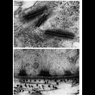
CIL:11209
NCBI Organism Classification
Phodopus
Biological Process
cell adhesion
Cellular Component
desmosome
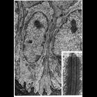
Upper panel, adjoining portions of two cells in the stratum spinosum of hamster cheek pouch epithelium shows desmosome junctions between two cells. Lower, a portion of the basal surface of a cell in ...
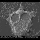
CIL:40624
NCBI Organism Classification
Homo sapiens
Biological Process
substrate-dependent cell migration
Cellular Component
cell surface
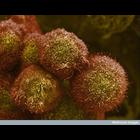
Scanning electron micrograph of breast cancer cells that have aggregated.
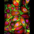
CIL:48102
NCBI Organism Classification
Homo sapiens
Biological Process
cell adhesion
Cellular Component
cytoskeleton
Cryopreserved human mammary epithelial cells were revived and stained for cytokeratin 18 (in red) to reveal the keratin-containing intermediate filaments found in the intracytoplasmic cytoskeleton of ...
CIL:11213
NCBI Organism Classification
Rattus
Biological Process
cell adhesion
Cellular Component
desmosome
Stratified squamous epithelium from the esophagus of the rat. Keratin filaments (tonofilaments, arrows) are sparse, and desmosomes are few (see inset), in the basal cell layer of this tissue. Figures...
CIL:39021
NCBI Organism Classification
none specified
Biological Process
cell-cell adhesion
Cellular Component
cell surface
Colorized scanning electron micrograph showing two lung cancer cells. These cells were grown using cell culture techniques.
CIL:39076
NCBI Organism Classification
none specified
Biological Process
cell-cell adhesion
Cellular Component
cell surface
A colorized scanning electron micrograph of pancreatic cancer cells grown in culture. See additional image at CIL: 39104.
CIL:39049
NCBI Organism Classification
none specified
Biological Process
substrate-dependent cell migration, cell attachment to substrate
Cellular Component
cell surface
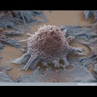
Colorized scanning electron micrograph of a lung cancer cell grown in culture.
CIL:24065
NCBI Organism Classification
Canis lupus familiaris
Biological Process
cell-cell adhesion
Cellular Component
adherens junction
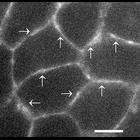
E-cadherin local dynamics were studied in mature junctions, that is, junctions engaged in adhesion for many hours, in which cadherin expression level is stable. After stable transfection with E-cadher...
CIL:24066
NCBI Organism Classification
Canis lupus familiaris
Biological Process
cell-cell adhesion
Cellular Component
adherens junction
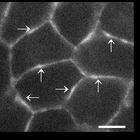
E-cadherin local dynamics were studied in mature junctions, that is, junctions engaged in adhesion for many hours, in which cadherin expression level is stable. After stable transfection with E-cadher...
« Previous
1
2
3
4
5
6
7
8
9
...
35
Next »
Results per page:
10
20
50
100