Alternate header for print version
Contributors
Help
Submit
Search
menu
Data sets
Videos
Latest data
Center for Research in Biological Systems
Basic Science Building, Room 1000
University of California, San Diego
9500 Gilman Drive
La Jolla, CA 92093-0608, USA
Voice
: (858) 534-0276
Fax
: (858) 534-7497
Email
: dorloff@ncmir.ucsd.edu
Search Results for
actin cytoskeleton
(1619 results)
CIL:35060
NCBI Organism Classification
none specified
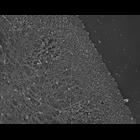
Biological Process
branching of actin filaments
Cellular Component
actin cytoskeleton
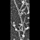
Electron micrograph of keratocyte or fibroblast lamellipodial actin network after unprotected extraction. All examples demonstrate frequent branching of actin filaments. Image corresponds to a singl...
CIL:35204
NCBI Organism Classification
Fundulus heteroclitus
Biological Process
gastrulation
Cellular Component
actin cytoskeleton
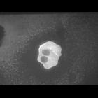
Individual deep cell moving by circus movements. Fundulus heteroclitus(killifish) embryo injected with GFP-actin plasmid at 1-cell stage, filmed during gastrulation. Image made available through Zebra...
CIL:34924
NCBI Organism Classification
Xenopus laevis
Biological Process
cellular localization
Cellular Component
lamellipodium
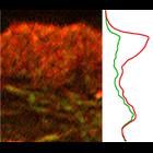
Localization of cross-linking proteins in fibroblast cytoskeleton. Fluorescence microscopy and corresponding intensity profiles of Xenopus fibroblast lamellipodia double stained with TRITC-phalloidin ...
CIL:7051
NCBI Organism Classification
Mus musculus
Biological Process
actin cytoskeleton organization
Cellular Component
actin cytoskeleton
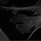
NIH 3T3 cell expressing EGFP-Lifeact (a small genetically expressed probe that binds to actin and is derived from the first 17 aa of Abp140) imaged with structured illumination. Structured illuminati...
CIL:38956
NCBI Organism Classification
none specified
Biological Process
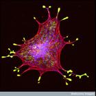
melanosome localization
Cellular Component
melanosome
Confocal micrograph of an isolated melanin-producing cell (a melanocyte) showing the melanosomes (vesicles that hold the melanin granules) in yellow, the actin in red and the microtubules in blue. The...
CIL:39061
NCBI Organism Classification
Bos taurus
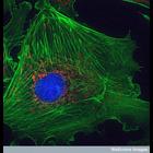
Biological Process
cytoskeleton organization
Cellular Component
mitochondrion
This image shows a bovine pulmonary artery endothelial cell. The pulmonary artery is the large vessel that takes deoxygenated blood cells from the heart to the lungs. These cells have been stained wit...
CIL:40810
NCBI Organism Classification
Rattus
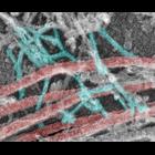
Biological Process
dendrite morphogenesis
Cellular Component
axon
Colorized transmission electron micrograph of a platinum replica showing the cytoskeletal organization of stubby dendritic spines in extracted hippocampal neurons after 14 DIV. This imge shows branche...
CIL:24808
NCBI Organism Classification
Xenopus laevis
Biological Process
cellular localization
Cellular Component
lamellipodium
Localization of XAC (Xenopus ADF/cofilin) in Xenopus keratocytes done with immuno-EM. A low mag view of the cell from which this high mag view is taken is shown in CIL 24807. Image corresponds to Fi...
CIL:39058
NCBI Organism Classification
none specified
Biological Process
neuron migration
Cellular Component
actin cytoskeleton
This confocal micrograph shows a dorsal root ganglion (DRG) explant. The dorsal root ganglion is a swelling on the dorsal roots of spinal nerves, which contains a cluster of cell bodies and synapses. ...
CIL:10113
NCBI Organism Classification
Rattus
Biological Process
developmental process
Cellular Component
cytoskeleton
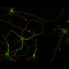
This multi-layer image shows the spatial relationship between filamentous actin (red) and microtubule array (green) in cultured hippocampal neurons, grown for 5 days in vitro. Actin staining (with rh...
« Previous
1
...
6
7
8
9
10
11
12
13
...
162
Next »
Results per page:
10
20
50
100