Alternate header for print version
Contributors
Help
Submit
Search
menu
Data sets
Videos
Latest data
Center for Research in Biological Systems
Basic Science Building, Room 1000
University of California, San Diego
9500 Gilman Drive
La Jolla, CA 92093-0608, USA
Voice
: (858) 534-0276
Fax
: (858) 534-7497
Email
: dorloff@ncmir.ucsd.edu
Search Results for
actin cytoskeleton
(1619 results)
CIL:24790
NCBI Organism Classification
none specified
Biological Process
branching of actin filaments
Cellular Component
lamellipodium
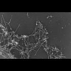
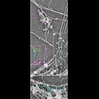
Improved visualization of actin filament branching in lamellipodia. EM of keratocyte or fibroblast lamellipodial actin network after cytochalasin D treatment (0.2 μM for 30 min or 0.5 μM for 10 min)...
CIL:35066
NCBI Organism Classification
none specified
Biological Process
branching of actin filaments
Cellular Component
actin cytoskeleton
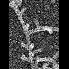
Electron micrograph of keratocyte or fibroblast lamellipodial actin network after unprotected extraction. All examples demonstrate frequent branching of actin filaments. Image corresponds to a singl...
CIL:34895
NCBI Organism Classification
none specified
Biological Process
branching of actin filaments
Cellular Component
actin cytoskeleton
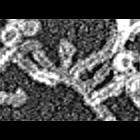
Improved visualization of actin filament branching in lamellipodia. EM of keratocyte or fibroblast lamellipodial actin network after cytochalasin D treatment (0.2 μM for 30 min or 0.5 μM for 10 min)...
CIL:34896
NCBI Organism Classification
none specified
Biological Process
branching of actin filaments
Cellular Component
actin cytoskeleton
Improved visualization of actin filament branching in lamellipodia. EM of keratocyte or fibroblast lamellipodial actin network after cytochalasin D treatment (0.2 μM for 30 min or 0.5 μM for 10 min)...
CIL:34901
NCBI Organism Classification
none specified
Biological Process
branching of actin filaments
Cellular Component
actin cytoskeleton
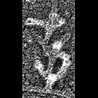
Improved visualization of actin filament branching in lamellipodia. EM of keratocyte or fibroblast lamellipodial actin network after cytochalasin D treatment (0.2 μM for 30 min or 0.5 μM for 10 min)...
CIL:31916
NCBI Organism Classification
Saccharomyces cerevisiae
Biological Process
receptor-mediated endocytosis
Cellular Component
endocytic vesicle
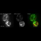
Localization of Alexa Fluor-594-α-factor-labeled endosomes (red), Abp1-GFP- (green) and Abp140-3GFP- (green) labeled endocytic vesicles. It was possible to distinguish endocytic vesicles from actin c...
CIL:40664
NCBI Organism Classification
Rattus
Biological Process
dendritic spine organization
Cellular Component
dendritic spine
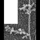
Platinum replica illustrating the cytoskeletal organization of dendritic spines from extracted 14 DIV neurons. Thin spines associate with dendrites at the base (bottom) and with axons by the head (top...
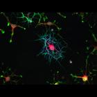
CIL:40358
NCBI Organism Classification
Rattus norvegicus
Biological Process
oligodendrocyte development
Cellular Component
actin cytoskeleton
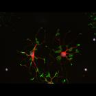
This fluorescent micrograph is part of an image group displaying the morphological changes associated with a developing oligodendrocyte. The progression of images starts with CIL:40358 where cells di...
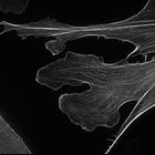
CIL:40360
NCBI Organism Classification
Rattus norvegicus
Biological Process
oligodendrocyte development
Cellular Component
actin cytoskeleton
This fluorescent micrograph is part of an image group displaying the morphological changes associated with a developing oligodendrocyte. The progression of images starts with CIL:40358 where cells di...
CIL:7053
NCBI Organism Classification
Mus musculus
Biological Process
actin cytoskeleton organization
Cellular Component
actin cytoskeleton
NIH 3T3 cell expressing EGFP-Lifeact (a small genetically expressed probe that binds to actin and is derived from the first 17 aa of Abp140) imaged with structured illumination. Structured illuminati...
« Previous
1
...
5
6
7
8
9
10
11
12
...
162
Next »
Results per page:
10
20
50
100