Alternate header for print version
Contributors
Help
Submit
Search
menu
Data sets
Videos
Latest data
Center for Research in Biological Systems
Basic Science Building, Room 1000
University of California, San Diego
9500 Gilman Drive
La Jolla, CA 92093-0608, USA
Voice
: (858) 534-0276
Fax
: (858) 534-7497
Email
: dorloff@ncmir.ucsd.edu
Search Results for
actin cytoskeleton
(1619 results)
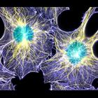
CIL:240
NCBI Organism Classification
none specified
Biological Process
none specified
Cellular Component
actin cytoskeleton
Two interphase cells with immunofluorescence labeling of actin filaments (purple), microtubules (yellow), and nuclei (green). This image won first place in the Nikon 2003 Small World photo competition...
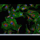
CIL:42155
NCBI Organism Classification
Mus musculus
Biological Process
dissemination
Cellular Component
plasma membrane
Representative time-lapse movie of a normal mouse mammary organoid in a 3D collagen I matrix. The cells are expressing td tomato as a membrane marker and eEGFP-actin using a keratin-14 promoter. Ima...
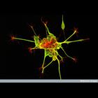
CIL:38974
NCBI Organism Classification
none specified
Biological Process
neuron migration
Cellular Component
growth cone
Dorsal root ganglion nerve cells stained to reveal the microtubules (green) and actin filaments (red). The axon shaft contains bundles of microtubules that give structural support and carry cargo (pro...
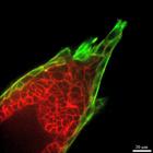
CIL:39059
NCBI Organism Classification
Bos taurus
Biological Process
cytoskeleton organization
Cellular Component
mitochondrion
This confocal image shows bovine pulmonary artery cells visualized with DAPI to highlight the nucleus (blue), MitoTracker Red CMXRos to stain mitochondria (red dots), and Alexafluor 488 phalloidin to ...
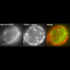
CIL:31922
NCBI Organism Classification
Saccharomyces cerevisiae
Biological Process
receptor-mediated endocytosis
Cellular Component
endocytic vesicle
Localization of Alexa Fluor-594-α-factor-labeled endosomes (center; red in merge) and Abp140-3GFP (left; green in merge). Abp140p binds F-actin and localizes to actin patches and cables. Endosome mov...
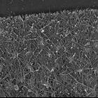
CIL:24786
NCBI Organism Classification
Xenopus laevis
Biological Process
branching of actin filaments
Cellular Component
lamellipodium
Multiple branching of actin filaments in lamellipodia of Xenopus keratocytes. This image shows an overview of the leading edge, and CIL 24787 shows enlargements of local regions of this platinum repli...
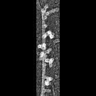
CIL:34894
NCBI Organism Classification
none specified
Biological Process
branching of actin filaments
Cellular Component
actin cytoskeleton
Improved visualization of actin filament branching in lamellipodia. EM of keratocyte or fibroblast lamellipodial actin network after cytochalasin D treatment (0.2 μM for 30 min or 0.5 μM for 10 min)...
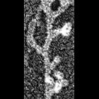
CIL:35062
NCBI Organism Classification
none specified
Biological Process
branching of actin filaments
Cellular Component
actin cytoskeleton
Electron micrograph of keratocyte or fibroblast lamellipodial actin network after unprotected extraction. All examples demonstrate frequent branching of actin filaments. Image corresponds to a singl...
CIL:35063
NCBI Organism Classification
none specified
Biological Process
branching of actin filaments
Cellular Component
actin cytoskeleton
Electron micrograph of keratocyte or fibroblast lamellipodial actin network after unprotected extraction. All examples demonstrate frequent branching of actin filaments. Image corresponds to a singl...
CIL:11844
NCBI Organism Classification
Potorous tridactylus
Biological Process
actin polymerization or depolymerization
Cellular Component
actin cytoskeleton
Actin dynamics in a Rac1(Q61L)-expressing PtK1 cell. Fast retrograde flow occurs in the lamellipodium and slow retrograde flow in the lamellum. The cell was microinjected with X-rhodamine-conjugated a...
« Previous
1
...
4
5
6
7
8
9
10
11
...
162
Next »
Results per page:
10
20
50
100